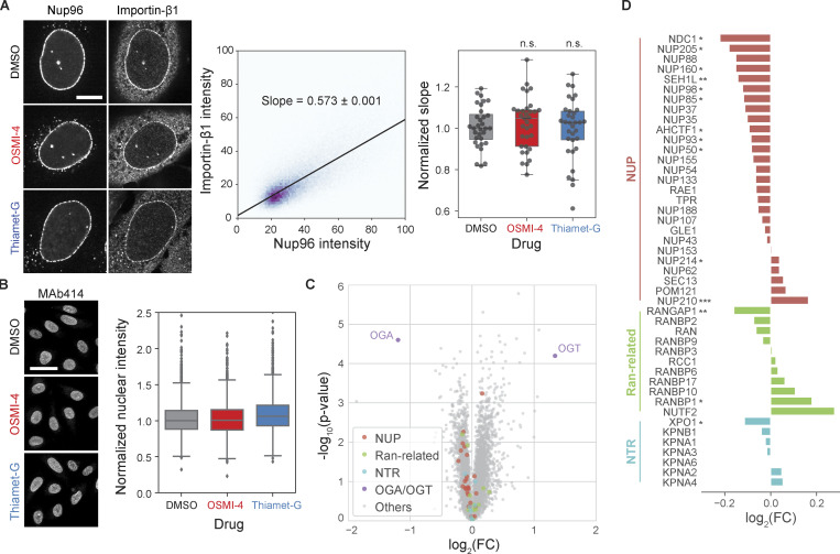
Figure S3.
O-GlcNAc perturbations do not affect importin-β1 localization to NPCs and protein levels of nuclear transport machinery. (A) Left: Importin-β1 and Nup96-GFP immunofluorescence images of cells treated with DMSO, 10 µM OSMI-4, or 10 µM Thiamet-G for 24 h. Middle: For each cell, a linear model (black line) was fitted to the pixel intensities of importin-β1 and Nup96 after background subtraction and thresholding. The slope of the linear model was used as a measure of the localization of importin-β1 at the NPCs. Right: The slopes for each drug condition. n > 31 cells for each condition. n.s., P > 0.5. (B) Left: MAb414 immunofluorescence images of cells treated with DMSO, 10 µM OSMI-4, or 10 µM Thiamet-G for 24 h. Right: Quantified mean nuclear intensity. n > 2,000 cells for each condition. For both A and B, data from the same batch were normalized to the median value of the DMSO condition. (C and D) Proteomics data from Martin et al. (2018) replotted to show the difference in the protein levels of nuclear transport machinery components between HEK293T cells treated with DMSO and those treated with 20 µM OSMI-4 for 24 h. FC, fold-change. *, P < 0.1; **, P < 0.01; ***, P < 0.001.

