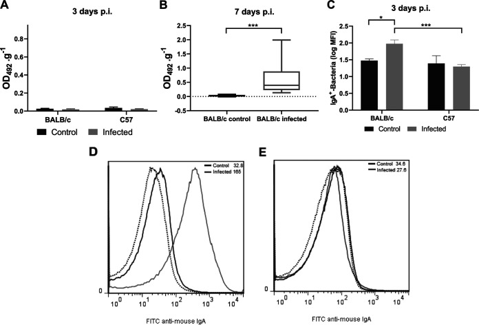
FIG 7.
Local humoral response during O157:H7 infection. Contents of the small and large intestine from noninfected (control) or infected mice were prepared as described in the Materials and Methods to determine anti-O157:H7 IgA in supernatants (1/2 dilution) by ELISA and IgA-coated bacteria in pellets by flow cytometry. Antibody levels were expressed as OD492.g−1. (A) Free anti-O157:H7 IgA in fecal supernatants at day 3 p.i. Each bar shows the mean ± SEM of 4 control mice and 6 infected mice for each mouse strain. (B) Free anti-O157:H7 IgA in fecal supernatants at day 7 p.i. The medians and interquartile ranges of 6 controls and 9 infected BALB/c mice are shown. Mann-Whitney test: ***, P< 0.001. (C) IgA-coated bacteria at day 3 p.i. Each bar shows the mean ± SEM of 4 control and 6 infected mice for each mouse strain. Two-way ANOVA test with Tukey’s posttest: *, P < 0.05; ***, P < 0.001. (D and E) Representative histograms of unstained samples (autofluorescence, dotted lines) and FITC anti-mouse IgA-stained samples from control (black lines) or infected (gray lines) BALB/c (D) and C57 (E) mice, assayed in parallel.

