Figure 1.
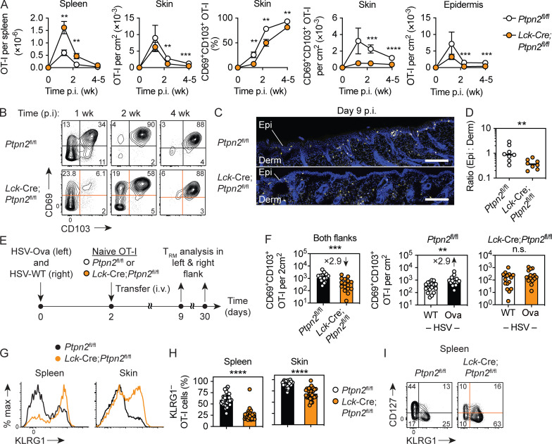
Absence of Ptpn2 promotes the generation of KLRG1+ effector cells and impairs TRM cell formation in response to HSV skin infection. (A–C) WT CD45.1+ mice were infected with HSV-Ova and 2 d later received naive Ptpn2fl/fl (white) or Lck-Cre;Ptpn2fl/fl (orange) CD45.2+ OT-I cells (5 × 10⁴ i.v.). (A) Enumeration of total and CD69+ CD103+ OT-I cells in spleen, skin, and epidermis by flow cytometry at indicated times post infection (p.i). Data pooled from n = 2 experiments with n = 8–9 mice/time point/group (mean ± SEM). (B) Analysis of CD69 and CD103 expression by OT-I cells in skin. (C and D) Immunofluorescence analysis of skin sections stained with Hoechst 33342 (blue) and anti-CD45.2 antibody to detect OT-I cells (yellow). Scale bars, 200 µm. Data in D were pooled from n = 2 experiments with n = 8 mice in total. Derm, dermis; Epi, epidermis. (E) WT CD45.1+ mice were infected with both HSV-Ova (left flank) and WT HSV (right flank) and 2 d later received congenic naive Ptpn2fl/fl (white) or Lck-Cre;Ptpn2fl/fl (orange) CD45.2+ OT-I cells (5 × 10⁴ i.v.). (F) Enumeration of CD69+ CD103+ OT-I TRM cells in skin 30 d p.i.; data were pooled from n = 3 experiments with n = 16–17 mice/group. (G–I) Analysis of KLRG1 and (I) CD127 expression by Ptpn2fl/fl and Lck-Cre;Ptpn2fl/fl OT-I cells in spleen and skin 9 d p.i.; data in H pooled from n = 7 experiments with n = 31 mice/group. Statistical significance (**, P < 0.01; ***, P < 0.001; ****, P < 0.0001) determined by Mann–Whitney test for individual time points. Derm, dermis; Epi, epidermis.

