Figure 2.
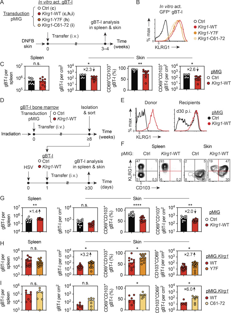
Forced KLRG1 expression impedes TRM cell formation in skin. (A) gBT-I cells were activated in vitro and transduced with a control vector (pMIG.Ctrl) or vectors coding for WT (pMIG.Klrg1.WT) or mutant (pMIG.Klrg1.Y7F; pMIG.Klrg1.C61-72) KLRG1. Sorted GFP+ cells were i.v. transferred (2.5 × 10⁶ in C; 1 × 10⁶ in H and I) into WT mice treated on flank skin with DNFB and gBT-I cells in spleen and skin analyzed 3–4 wk later. (B) Analysis of KLRG1 expression by transduced GFP+ gBT-I cells before transfer. (C) Enumeration of total and CD69+ CD103+ gBT-I cells in spleen and skin 4 wk after transfer (pMIG.Ctrl, white; pMIG.Klrg1.WT, red). Data pooled from n = 2 experiments with n = 10 mice per group. (D–G) Bone marrow cells from gBT-I mice were transduced with pMIG.Ctrl or pMIG.Klrg1.WT and transferred into lethally irradiated WT mice. Following reconstitution, GFPhi KLRG1hi Vα2+ CD8+ gBT-I cells were isolated, sorted, and transferred (2–10 × 104 i.v.) into congenic mice infected with HSV-WT 1 d earlier. (E) Analysis of KLRG1 expression by GFP+ gBT-I cells from donor and recipient mice (pMIG.Ctrl, black; pMIG.Klrg1.WT, red). (F and G) Analysis of KLRG1 and CD103 expression by gBT-I cells in spleen and skin (F; numbers indicate percentage in each quadrant) and enumeration of total and CD69+ CD103+ gBT-I cells 4 wk after infection (G). Data pooled from n = 2 experiments with n = 12 mice/group. Ctrl, control. (H and I) gBT-I cells were transduced with pMIG.Klrg1.WT (red), pMIG.Klrg1.Y7F (orange), or pMIG.Klrg1.C61-72 (light orange) and transferred into DNFB-treated recipients as described in A. Enumeration of total and CD69+CD103+ GFP+ gBT-I cells in spleen and skin 3 wk after transfer. Data pooled from n = 4 experiments with n = 15–17 mice/group (H) or from n = 2 experiments with n = 7 mice/group (I). Statistical significance (*, P < 0.05; **, P < 0.01; ****, P < 0.0001) determined by Mann–Whitney test.

