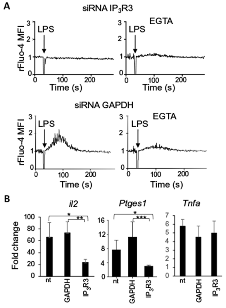
Fig. 3. IP3R3 channels are required for LPS-induced Ca2+ mobilization and NFAT activation in DCs.

(A) Ca2+ transients induced by LPS in D1 cells, 48 hours after the knockdown of IP3R3 with specific siRNAs in the presence or absence of the extracellular calcium chelator EGTA. Knockdown of GAPDH with specific siRNA was used as negative control. Arrows indicate the time (30 s) of LPS administration. Ca2+ fluctuations were evaluated by FACS on bulk populations as changes in the Fluo-4 fluorescence, in response to the stimuli, and were normalized to the value during the 30 s before LPS addition (rFluo-4 MFI). Traces are representative of three independent experiments. (B) Real-time polymerase chain reaction (PCR) analysis of the increase in il2, Ptges1, and Tnfa expression after 4 hours of LPS treatment in D1 cells transfected or not (nt) with siRNAs specific for GAPDH (control) or IP3R3. Values indicate the mean ± SEM from six independent experiments. Statistical significance was determined with one-way analysis of variance, followed by Tukey’s multiple comparisons test, *P < 0.05, **P < 0.01 and ***P < 0.001.
