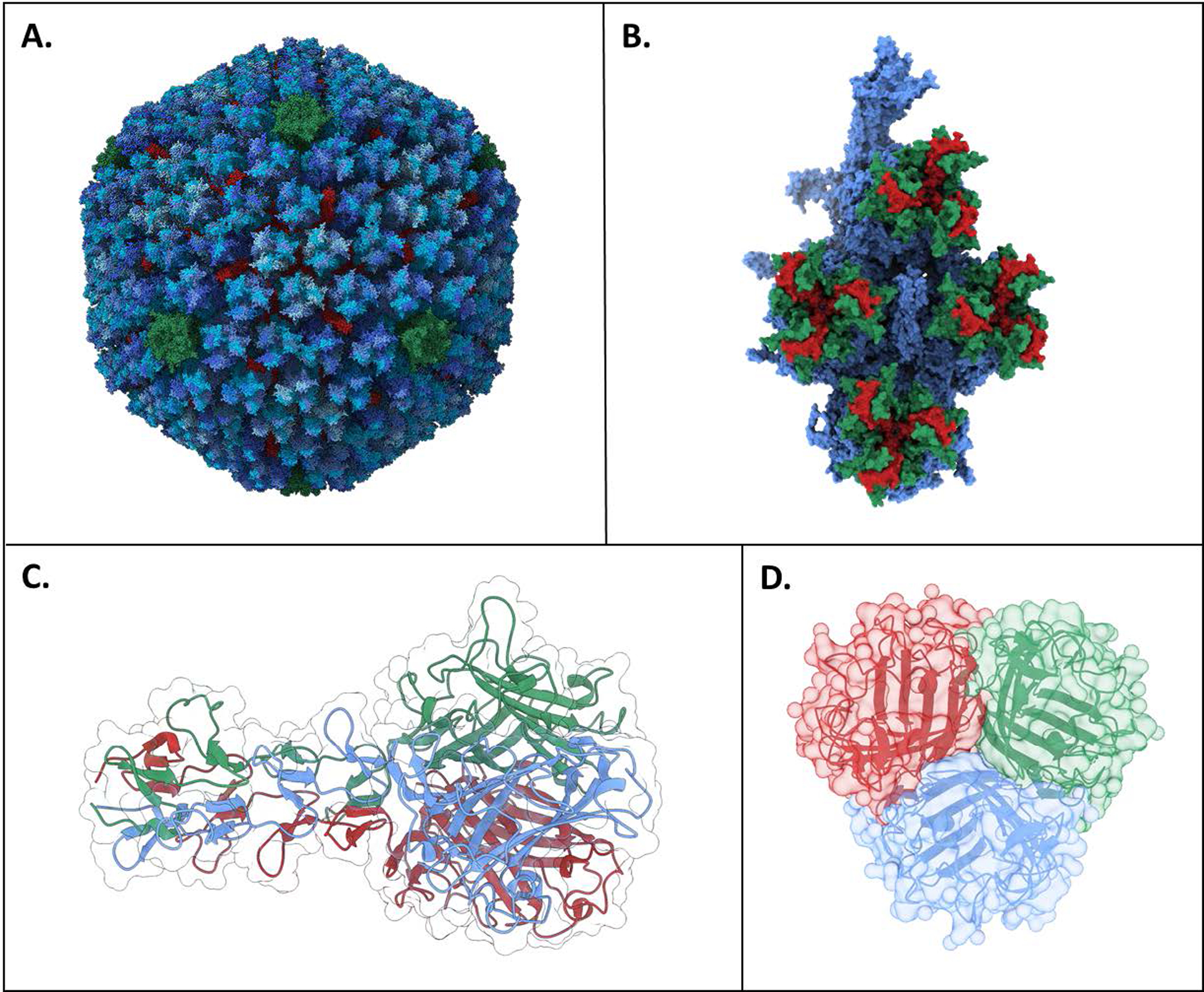
Figure 2:

Adenovirus structure. A) Adenovirus capsid structure. The various hexon protein chains are colored in blues, penton protein in green, and hexon-interlacing protein in red. Trimer fiber inserts into green penton protein center (not shown). B) Hexon monomeric unit. Hypervariable loop 1 is shown on green and hypervariable loop 2 is shown in red. Alignment of hypervariable regions determined from [50] C) Side view of fiber knob from Ad2 (structurally similar to Ad5 and four shaft repeat motifs. D) Top view of fiber Ad5 fiber knob showing trimeric symmetry. PDB IDs: 6B1T (A, B) [51], 1QIU (C) [52], and 6HCN (D) [53]. Rendered with UCSF ChimeraX [25]. Print in color.
