Fig. 2.

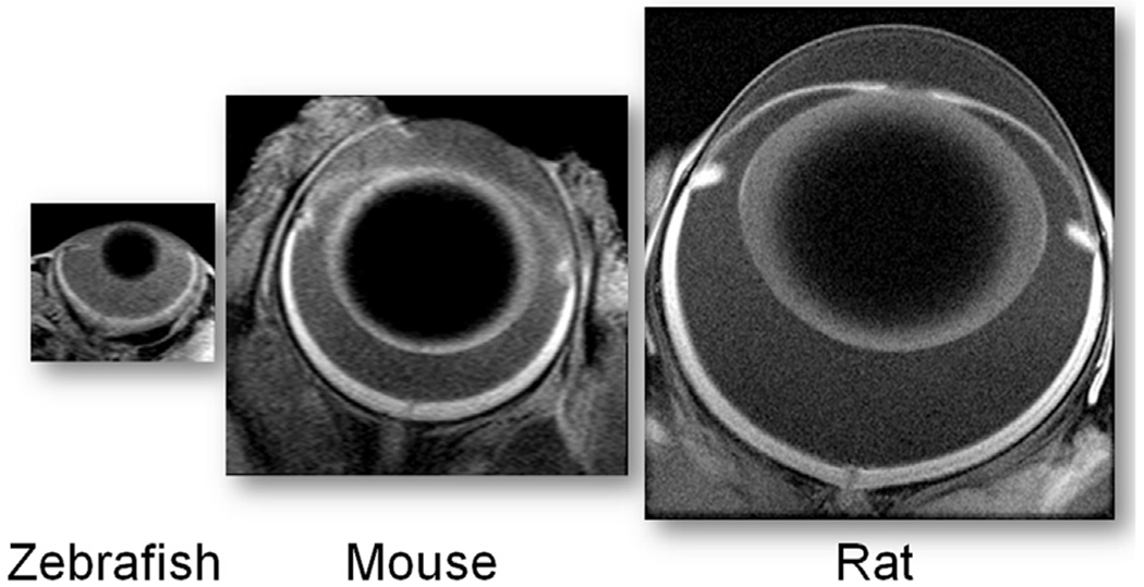
Representative MRI images (axial resolution 23.4 μm) of three common laboratory models acquired using established protocols (Berkowitz et al., 2014; Bissig et al., 2013); the images are proportionately sized based on ocular anatomy.

Representative MRI images (axial resolution 23.4 μm) of three common laboratory models acquired using established protocols (Berkowitz et al., 2014; Bissig et al., 2013); the images are proportionately sized based on ocular anatomy.