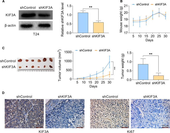
Figure 4.

Knockdown of KIF3A impaired tumor growth of bladder cancer cells in vitro. (A–C) T24 cells were infected with KIF3A or control shRNA lentivirus, and subsequently implanted into nude mice. After 2 weeks, tumors were isolated, and volume was calculated every 5 days. After 30 days, all tumors were isolated (n = 6 in each group). Tumor growth curves were calculated based on the average volume of six tumors for each group. (A) Immunoblot assays confirmed the efficiently silenced KIF3A expression in tumor tissues from KIF3A‐depleted mice. (B) Weight of mice from control or KIF3A depletion groups. (C) Representative photographs of tumors, the growth curve and tumor weight were exhibited, respectively. (D) Immunohistochemical assays indicated the expression levels of KIF3A and Ki67 in control or KIF3A knockdown tumor tissues isolated from mice (mean ± SEM, **P < 0.01). Scale bars indicate 50 μm. Student’s t‐test was used for statistical comparisons, and three independent replicates were performed.
