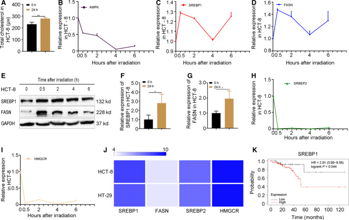
Fig. 2.

Radiation exposure precipitates cholesterol synthesis through SREBP1/FASN signaling in CRC cells. (A) The level of cholesterol in HCT‐8 cells at 24 h after 6 Gy γ‐ray irradiation. (B) The dynamic expression of AMPK was examined by qRT‐PCR at 0, 0.5, 2, 4 and 6 h after 6 Gy γ‐ray irradiation in HCT‐8 cells. (C–E) The dynamic expression of SREBP1 and FASN was examined by qRT‐PCR at 0, 0.5, 2, 4 and 6 h after 6 Gy γ‐ray irradiation in HCT‐8 cells. (F, G) The expression of SREBP1 and FASN was examined by qRT‐PCR at 24 h after 6 Gy γ‐ray irradiation in HCT‐8 cells. (H, I) The dynamic expression of SREBP2 and HMGCR was examined by western blotting at 0, 0.5, 2, 4 and 6 h after 6 Gy γ‐ray irradiation in HCT‐8 cells. (J) The basal expression of SREBP1/FASN and SREBP2/HMGCR in HCT‐8 and HT‐29 cell lines was analyzed by ccle (https://portals.broadinstitute.org/ccle). Color depth represents the intensity of expression. (K) Kaplan–Meier analysis of the overall survival rate of rectum adenocarcinoma patients (http://kmplot.com/analysis). Data are shown as the mean ± SD. GAPDH was used as a loading control. Statistical significance: **P < 0.01; ***P < 0.001, Student’s t‐test.
