
Fig. 4.
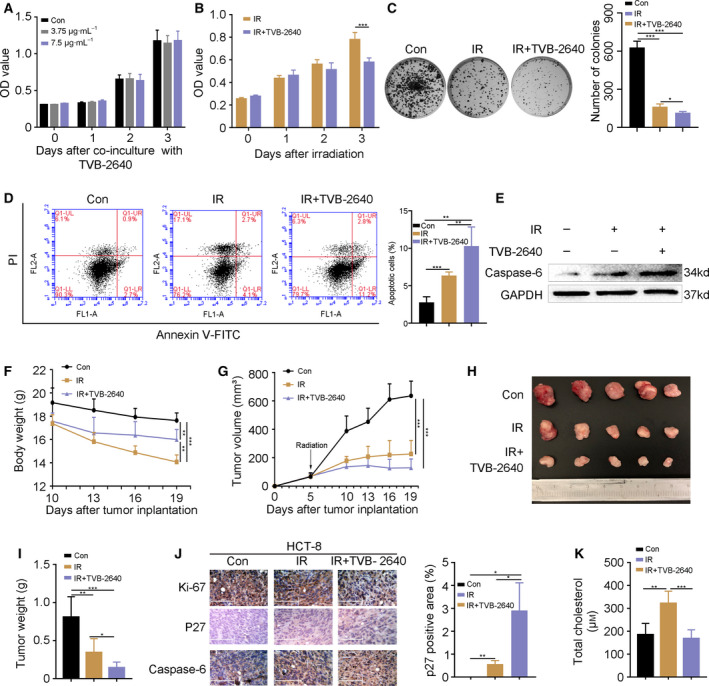
FASN inhibitor accelerates CRC cell death following radiation exposure. (A) A CCK‐8 assay showed the cell viability of HCT‐8 cells treated with TVB‐2640. (B, C) The proliferation of HCT‐8 cells with or without TVB‐2640 treatment was assessed by CCK‐8 and colony formation assays following radiation challenge. (D) Flow cytometric analysis showed the apoptosis in HCT‐8 cells treated with TVB‐2640 at 24 h after 6 Gy γ‐ray irradiation. (E) The expression of caspase‐6 was examined by western blotting in HCT‐8 cells treated with TVB‐2640 at 24 h after 6 Gy γ‐ray irradiation. (F) The body weight of nude mice. (G–I) Growth curve, photograph and weight of HCT‐8 tumors from nude mice. (J) The expression of Ki‐67, p27 and caspase‐6 was detected by immunohistochemistry staining in HCT‐8 tumors from nude mice. Scale bars = 125 μm. The p27 positive area was analyzed using image‐pro plus. (K) The level of cholesterol in HCT‐8 tumor from nude mice. Data are shown as the mean ± SD. GAPDH was used as a loading control. Statistical significance: *P < 0.05; **P < 0.01; ***P < 0.001, Student’s t‐test.