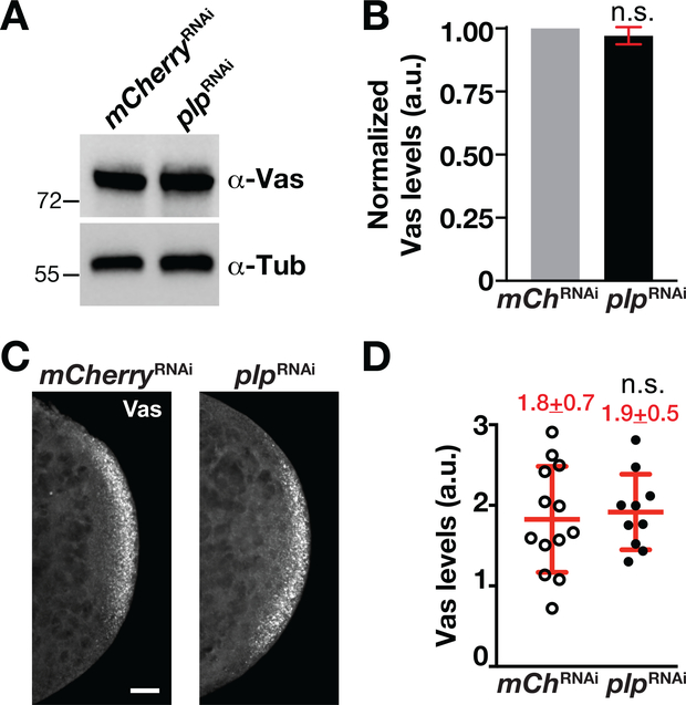
FIGURE 2.
PLP is dispensable for Vas localization to posterior cortex. (a) Western blot analysis of Vas expression from 0–1.5 hr mChRNAi (mCherryRNAi) and plpRNAi embryo extracts. Anti-α-Tub antibodies were used for normalization. (b) Densitometry quantification of Vas levels from western blot analysis. Vas expression values are relative to the α-Tub loading control and normalized to the mCherryRNAi. Error bars show ± SD from three replicates. a.u., arbitrary units; n.s., not significant by Student’s t test. (c). Images show maximum-intensity projections of 0–1.5 hr embryos of the indicated genotypes stained with anti-Vas (gray scale). Bar, 10 μm. (d) Quantification of Vas levels measured within a region-of-interest at the posterior pole. Each data point indicates a single measurement from one embryo, mCherryRNAi (N = 13 embryos) and plpRNAi (N = 10 embryos). Mean ± SD Vas levels are displayed (red). n.s., not significant by Student’s t test. The experiment was performed twice with similar results. PLP, Pericentrin-like protein

