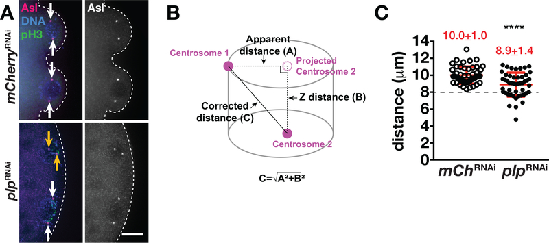
FIGURE 4.
PLP is required for centrosome separation. (a) Images show maximum-intensity projections of prophase-stage NC 10 embryos of the indicated genotypes stained with anti-pH3 (green) to label mitotic nuclei and anti-Asl (magenta) to label the centrosomes. DAPI labels the nuclei (blue). Arrows mark complete (white) or incomplete (orange) centrosome separation. Dashed lines outline the posterior cortex. Bar, 10 μm. (b) Cartoon depicts the distance between a pair of centrosomes. A, represents the apparent distance between centrosomes from a projected image. B, represents the Z-distance between the two centrosomes. C, represents the true distance, as calculated by the Pythagorean theorem. (c) Quantification shows centrosome distance during prophase NC 10 in mCherryRNAi (N = 49 events from 12 embryos) and in plpRNAi (N = 48 events from 10 embryos). ****p < .0001. Mean ± SD are displayed (red). Dashed line indicates 8 μm. The experiment was performed twice with similar results. NC, nuclear cycle; PLP, Pericentrin-like protein

