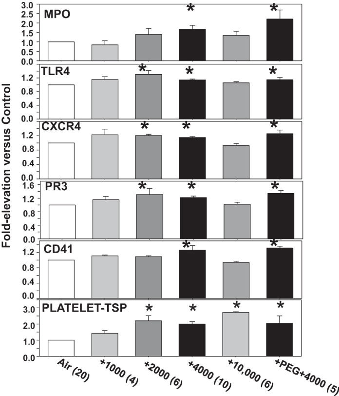
Fig. 2.

Neutrophil and platelet activation. Surface proteins on neutrophils and platelets were quantified in mice manipulated as described in the caption for Fig. 1. Frames 1–5: analyses of Ly6G-positive cells (neutrophils) where surface expression was evaluated for myeloperoxidase (MPO), Toll-like receptor 4 (TLR4), CXC chemokine receptor 4 (CXCR4), proteinase 3 (PR3), and presence of platelet-specific CD41 as reflecting platelet-neutrophil interactions. Frame 6: elevation in surface expression of thrombospondin-1 (TSP) on platelets, which were identified as CD41-positive, between 3 and 5 µm diameter and annexin V-negative. Data are means ± SE; n is shown for each sample. *P < 0.05, significantly different from control by ANOVA.
