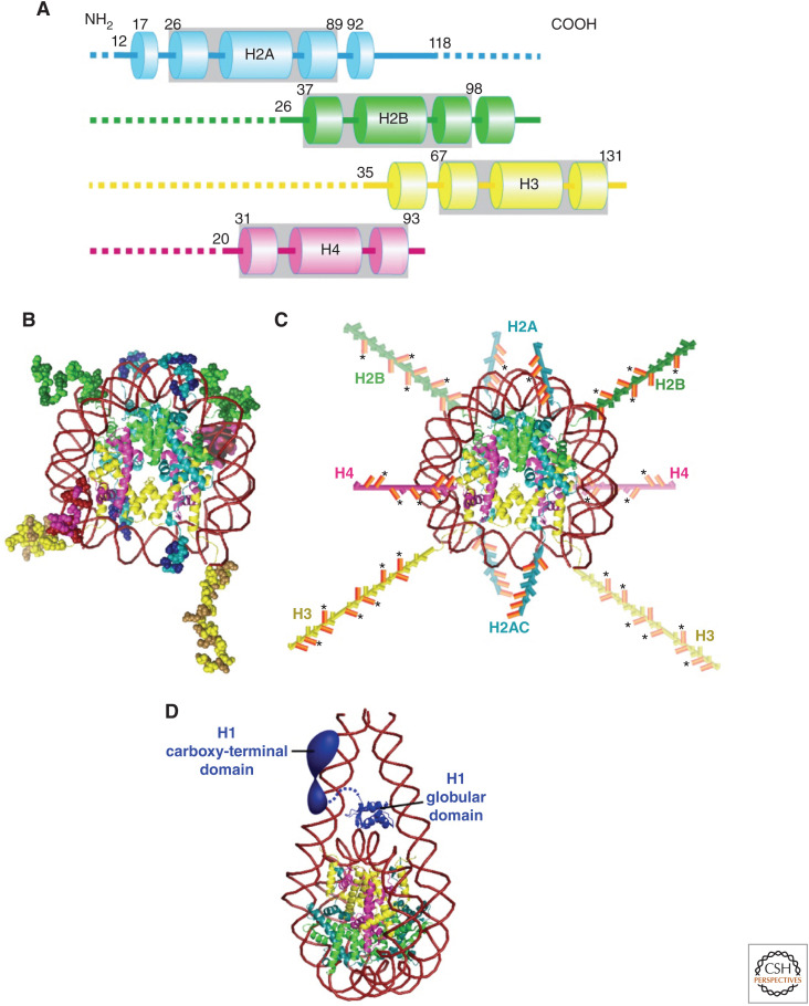
Figure 2.
Structures of core histones and nucleosome. (A) Schematic of the core histone structures; histone-fold domains are enclosed by gray boxes, tail domains are denoted by dashed lines, and α-helices are shown by columns. The positions of residues flanking the histone fold in each protein are shown. The approximate residue at the end of the tail domain nearest the histone fold is also shown. (B) The nucleosome structure at 1.9 Å resolution (Davey et al. 2002). Note that histone colors correspond to A. Amino acid residues of histone tails (intrinsically disordered regions [IDRs]) are shown as ball models. Basic residues (lysines and arginines) in the tail domains are highlighted with a darker color. (C) Amino-terminal tail domains in B are extended away from the nucleosome core. H2A also has a carboxy-terminal tail. Basic residues (lysines and arginines) in the tail domains are colored in orange. An asterisk indicates sites of lysine acetylation. (Illustrations are based on data in Wolffe and Hayes 1999 and Pepenella et al. 2014a.) (D) Structural model of a nucleosome with a linker histone H1. (The model in D was created from PDB data in Bednar et al. 2017.)

