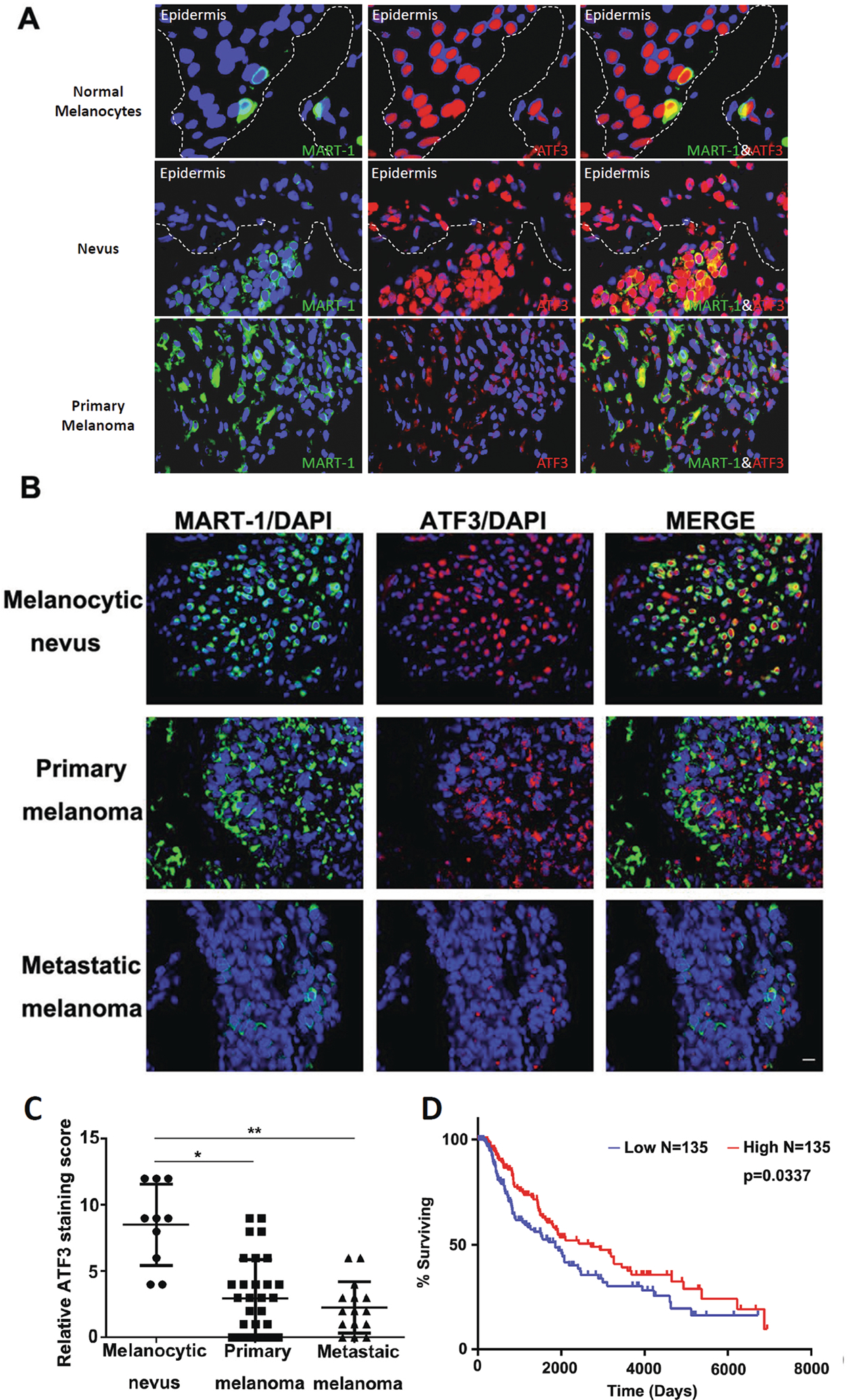
Figure 1. ATF3 expression decreased in melanoma.

(A and B) Representative IF images of double staining of ATF-3 (red) and MART-1 (green) in human melanocytic specimens and tissue microarray, respectively. (A). Strong nuclear positivity of ATF-3 (red) present in normal melanocytes (40x) and benign nevi (20x), while melanoma showed marked decrease (20x) in the representative images. (B). Representative IF images of tissue microarray of 72 patient cases of melanocytic lesions, which included benign nevi (n=10), primary cutaneous melanomas (n=38) and metastatic melanomas (n=24) by IF are shown (10x) and revealed significant decrease of ATF-3 nuclear positivity (red) with progression from nevi to primary melanoma (p<0.01), with an apparent qualitative diminution in nuclear staining seen in metastatic melanoma when compared to primary melanomas (p<0.05) (C). Kaplan-Meier survival curves on the ATF3 level in the TCGA skin cutaneous melanoma database (D). Data are presented as the mean ± standard deviation, *P < 0.05; **P < 0.01.
