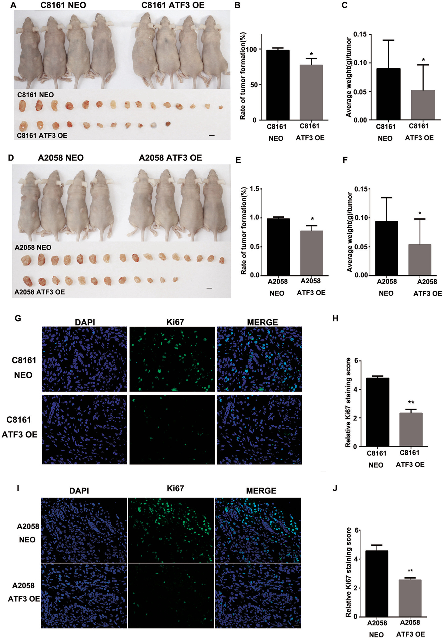
Figure 3. ATF3 over-expression inhibit melanoma tumor formation and growth in vivo.

(A) The representative of images of mice at 3 wk after subcutaneously injection of either C8161 NEO and C8161 ATF3 OE cells, and each tumor was collected shown in the lower panel. (B) The percentage of C8161 NEO and C8161ATF3 OE tumor formation and (C) average weight of each tumor was quantified. (D) Representative images of mice at 3 wk after subcutaneously injection of either A2058 NEO and A2058 ATF3 OE cells, and each tumor was collected shown in the lower panel. (E) The percentage of A2058 NEO and A2058 OE tumor formation and (F) average weight of each tumor was quantified. (G) Representative images of Ki67 immunofluorescence staining in C8161 NEO and C8161 ATF3 OE melanoma tumors and (H) summarized staining score of Ki67 expression. (I) Representative images of Ki67 immunofluorescence staining in A2058 NEO and A2058 ATF3 OE melanoma tumors and (J) summarized staining score of Ki67 expression. Data are presented as the mean ± standard deviation, *P<0.05; **P < 0.01.
