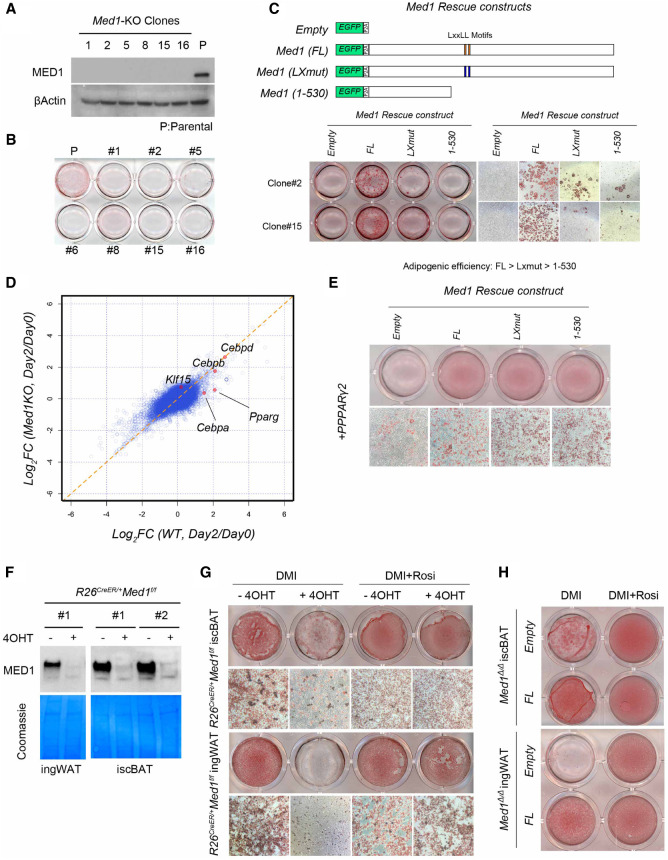
Figure 2.
MED1 is required for adipocyte differentiation in culture. (A) Immunoblot of lysates prepared from established 3T3-L1 clones, showing loss of MED1. β-Actin was used as a loading control. Parental 3T3-L1 cells were used as control. (B) Plate image of Oil Red O staining after 6 d of differentiation of parental and Med1-KO 3T3-L1 cells. None of the KO clones showed successful differentiation. (C) Lentiviral transduction of empty (eGFP only) vector or different forms of MED1 with the eGFP-P2A-construct. Note that only the full length restores efficient differentiation in the absence of Rosi. (D) Analysis of cDNA microarray data obtained from parental (WT) and pooled Med1-KO clones at day 0 and day 2 of differentiation. Ratio of expression levels at day 2 over day 0 is plotted for each condition. Classical adipogenic transcription factors are plotted in red dots. Note that Pparg and Cebpa are uninduced in KO conditions. (E) Ectopic PPARγ2 (introduced by retrovirus) rescue of Med1-KO 3T3-L1 clones with different forms of MED1. Note that efficient lipid accumulation does not occur in the absence of MED1. (F) Immunoblot of preadipocyte lysates. Efficient depletion of MED1 is seen only in 4OHT-treated (+) and not in vehicle-treated (−) cultures. Coomassie staining of corresponding membrane is shown as a loading control. (G) Oil Red O staining of differentiated brown and white preadipocytes pretreated with vehicle or 4OHT. (H) Oil Red O staining of differentiated brown and white preadipocytes pretreated with 4OHT and then transduced with lentivirus expressing empty (eGFP) or eGFP-P2A-MED1 constructs.

