Figure 3.
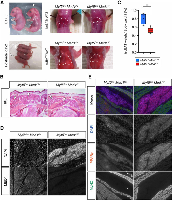
Brown adipose tissue is formed in Myf5CreMed1f/f mice. (A) Macroscopic examination of BAT of Myf5CreMed1f/w and Myf5CreMed1f/f mice at perinatal (E17.5 and P1) stages. (Left panel) White arrows indicate the Myf5CreMed1f/f mice. BAT is present at the upper back of the Myf5CreMed1f/f mice. (B) H&E staining of thoracic transversal cryosection of E17.5 Myf5CreMed1f/w and Myf5CreMed1f/f mice. Arrowheads indicate the location of BAT. (C) Percentage of BAT weight over body weight of Myf5CreMed1f/w (n = 4) and Myf5CreMed1f/f (n = 5) mice at P1 (mean ± SD, unpaired t-test, P < 0.005). (D) Immunohistochemistry of MED1 in E17.5 BAT regions. Absence of MED1 in Myf5CreMed1f/f mice is shown. Scale bars, 100 μm. (E) Immunohistochemistry of PPARγ (BAT marker) and MyHC (skeletal muscle) in E17.5 embryos. Scale bars, 100 μm.

