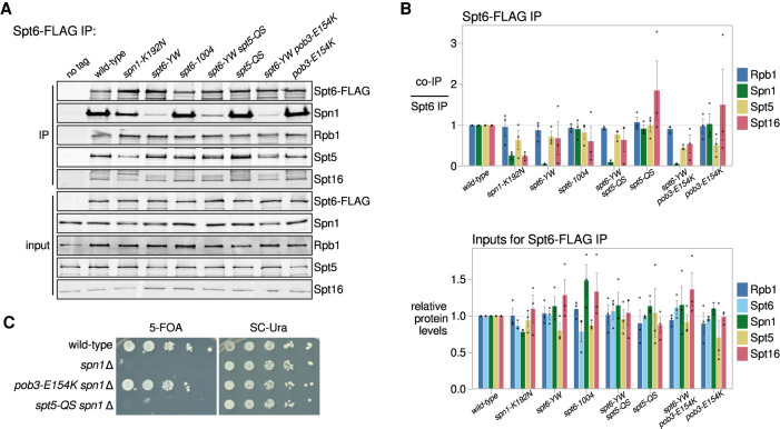
Figure 4.
The pob3-E154K and spt5-QS suppressors do not restore the Spt6-Spn1 interaction. (A) Western blots for Spt6-FLAG co-IP analysis, as in Figure 1A. Spt5 and Spt16 were detected using their respective polyclonal antibodies. (B) Quantification of Spt6-FLAG co-IP experiments (top) and inputs (bottom). Error bars indicate the mean ± standard error of the relative Western blot signal from the replicates shown. The co-IP signal was normalized to the Spt6-FLAG pull-down signal. (C) Assay for the ability of pob3-E154K or spt5-QS to suppress spn1Δ inviability. Growth in the presence of 5-fluoroorotic acid (5-FOA) indicates viability after the loss of a SPN1-URA3 plasmid as the sole source of Spn1. Strains were grown to saturation in YPD, serially diluted 10-fold, and spotted for growth on the indicated media.

