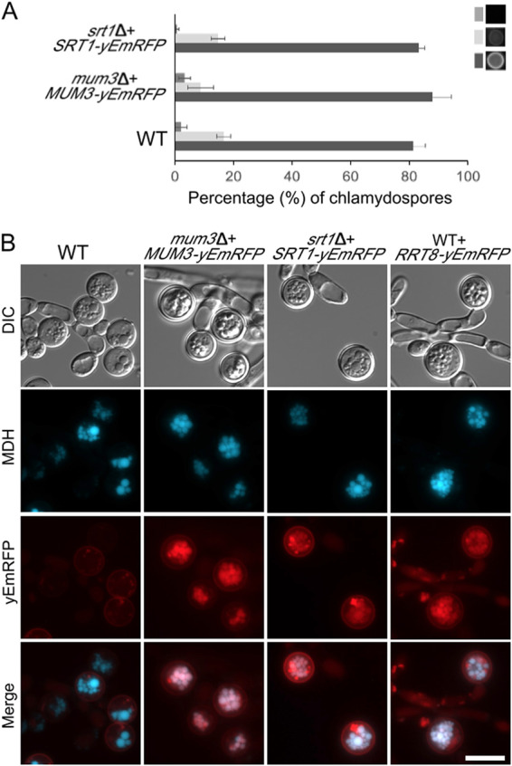
FIG 8.

Localization of Cda2, Mum3, Rrt8, and Srt1 in chlamydospores. (A) Eosin Y staining of chlamydospores in WT (CD1465) mum3Δ MUM3-yEMRFP (BEM20) and srt1Δ SRT1-yEmRFP (BEM22) strains was quantified as in Fig. 4B. (B) WT (Cd1456) cells expressing no RFP fusion or strains expressing different MUM3-, SRT1-, or RRT8-yEmRFP fusions (BEM20, -21, or -22) were grown on SGlycerol medium, stained with MDH, and visualized through both BFP and RFP filters. Scale bar, 10 μm.
