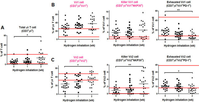
Figure 3.
Immunoassay of the gamma delta (γδ) T cell subsets before and after hydrogen inhalation in non-small cell lung cancer patients.
Note: (A) Change in the number of γδ T cells. (B) Test results of the Vδ1 subsets. (C) Test results of the Vδ2 subsets. The parallel red long lines in the figure represent the normal range, the black short lines represent the average value at each time point, and the pink cell names represent the abnormal indicators before hydrogen treatment. Data were analyzed by repeated measures analysis of variance followed by Bonferroni’s multiple comparison test. *P < 0.05, **P < 0.01, ***P < 0.001. NK: Natural killer ; PD-1: programmed cell death protein 1.

