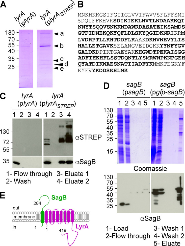
FIG 1.

SagB copurifies with LyrA. (A) Coomassie stain of desthiobiotin eluates following affinity purification over Strep-Tactin resin. Samples loaded on the column were enriched for membrane proteins by DDM solubilization of bacterial extracts from lyrA(plyrA) and lyrA(plyrAstrep) cultures, respectively. Arrowheads labeled a through e indicate bands excised for LC-MS/MS analysis. (B) The amino acid sequence of SagB with peptides identified by LC-MS/MS shown in bold. (C) Strep-Tactin purification of samples as described for panel A. The flowthrough, wash, and elution fractions (lanes 1 to 4) were analyzed by immunoblotting (αSagB and αStrep antibodies). (D) DDM fractions prepared from S. aureus overproducing SagB or GFP-SagB were purified over GFP-trap agarose. Aliquots of material loaded on the columns, flowthrough, wash, and eluates (lanes 1 to 5) were separated by SDS-PAGE and stained with Coomassie or analyzed by immunoblotting (αSagB antibodies). (E) Depiction of LyrA and SagB in the membrane. Numbers to the left of gels and blots indicate molecular weight markers in kilodaltons.
