FIG 1.
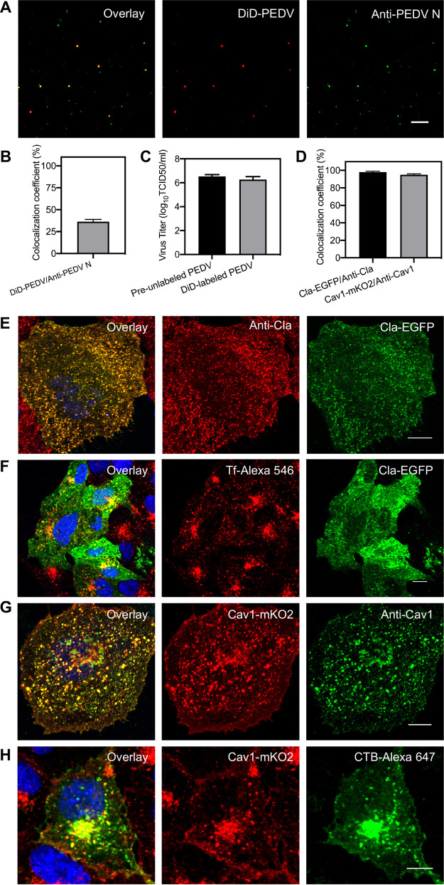
Characterization of DiD-labeled PEDVs and fluorescent protein-labeled endocytic structures. (A) Fluorescence images of DiD-labeled and immunostained PEDVs. (B) The efficiency of viral labeling by DiD dyes evaluated by the colocalization of DiD signal and anti-PEDV N protein signal. (C) Infectivity assay of preunlabeled PEDVs and DiD-labeled PEDVs measured by TCID50; the average of logarithms of TCID50 values in 3 independent experiments of preunlabeled PEDVs was 6.53 and that of DiD-labeled PEDVs was 6.26. (D) Colocalization analysis of discrete fluorescent protein signal (Cla-EGFP) and immunofluorescence signal (anti-Cla) of clathrin structures and fluorescent protein signal (Cav1-mKO2) and immunofluorescence signal (anti-Cav1) of caveolae structures, respectively. (E) Representative immunofluorescence images of clathrin-coated structures in Vero CCL81 cells. (F) Uptake of Tf in Vero-CCL81 cells expressing Cla-EGFP. (G) Representative immunofluorescence images of caveolae structures in Vero-CCL81. (H) Uptake of CTB subunit in Vero-CCL81 cells expressing Cav1-mKO2. In all panels, data shown are mean results ± standard deviation (SD) of three independent experiments, and white bars indicate 10 μm.

