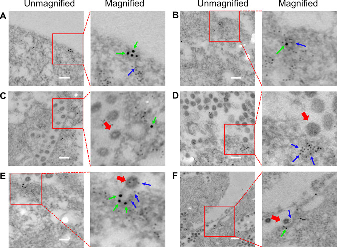
FIG 7.
Clathrin and caveolin-1 double immunogold electron microscopy of PEDV entry pathway. (A) Distinct regions of CCPs and caveolae on cell membrane. (B) Clathrin and caveolin-1 double-positive invagination on cell membrane. (C) CME of PEDV. (D) CavME of PEDV. (E, F) C3ME of PEDV. Clathrin was labeled with 10-nm immunogold, as indicated by the green arrow; caveolin-1 was labeled with 4-nm immunogold, as indicated by the blue arrow; and PEDV was indicated by the red arrow. Scale bar, 200 nm.

