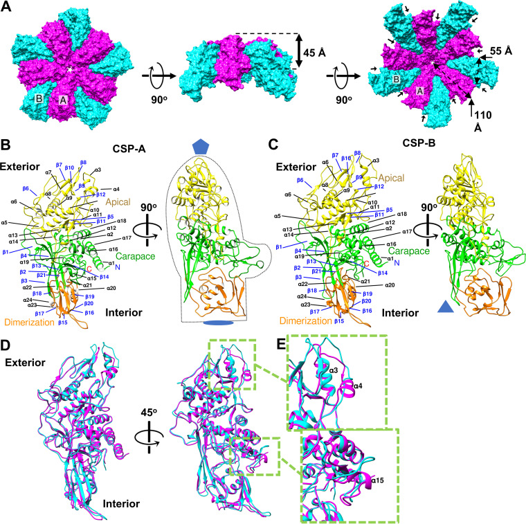
FIG 2.
Characterization of the capsid shell protein. (A) Surface diagrams of decamers subunits with CSP-As (magenta) nearest the I5 vertex and surrounded by CSP-Bs (cyan) viewed from the exterior (left), side (middle), and interior (right). Approximate measurements of the conformer dimensions are overlaid in Å. Stabilizing protrusions are labeled with black pointers. (B and C) Orthogonal views of A and B conformers (CSP-A and CSP-B), respectively, with secondary structural elements labeled, symbols indicating sites of icosahedral symmetry, and mitten shape provided to enhance visualization. Colored: carapace, green; apical, yellow; dimerization domain, orange. (D) Ribbon diagram of CSP-A (magenta) and CSP-B (cyan) superimposed upon one another. (E) Close-up view of sites of structural discrepancies between conformers display on winding of α4 (top) and α15 (bottom) from CSP-A to CSP-B.

