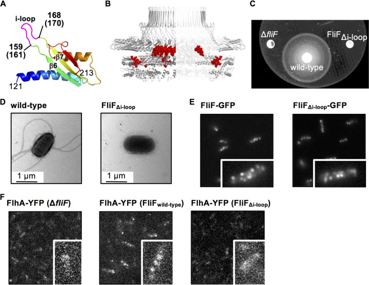
FIG 3.
Effect of the deletion of the loop between β4 and β5 (i-loop). (A) Structure of D2 of Aa-FliF. The i-loop is highlighted in magenta. The corresponding residue numbers of St-FliF are shown in parentheses. (B) The i-loops (red spheres) in the ring model. (C) Swimming motility of Salmonella wild-type and FliFΔi-loop mutant cells in the soft agar plate. The plates were incubated at 30°C for 9 h. (D) Electron micrographs of a Salmonella wild-type cell and a FliFΔi-loop mutant cell. (E) Subcellular localization of St-FliF-GFP and St-FliFΔi-loop-GFP. (F) Subcellular localization of FlhA-YFP in the fliF null mutant, the wild type, and the FliFΔi-loop mutant. Magnified views of single cells are shown in the lower right of each image in E and F.

