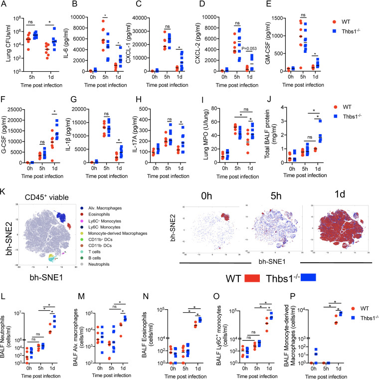
FIG 1.
Thrombospondin-1 (TSP-1) prevents excessive inflammatory cell recruitment by restraining the production of cytokines and chemokines in the lungs during P. aeruginosa infection. TSP-1-deficient (Thbs1−/−) and WT mice were intratracheally (i.t.) inoculated with P. aeruginosa at an inoculum of 106 CFU. (A) Lung bacterial burden (CFU/ml) was measured at 5 h postinfection (hpi) and at 1 day postinfection (dpi). In parallel, (B) IL-6, (C) CXCL-1, (D) CXCL-2, (E) GM-CSF, (F) G-CSF, (G) IL-1β, (H) IL-17A, and (I) myeloperoxidase (MPO) activity were measured in lung tissue homogenates after 5 hpi and 1 dpi. (J) Total bronchoalveolar lavage fluid (BALF) protein content was measured after 5 hpi and 1 dpi. (K) Immunophenotyping of BALF leukocytes was analyzed by the unbiased Barnes-Hut modification of t-SNE (bh-SNE) method using live CD45+ cells from WT and Thbs1−/− mouse samples at 0 h, 5 h, and 1 day postinfection. (Left) Clusters of leukocyte subsets based upon expression level of surface markers. (Right) Kinetics of leukocyte subsets in BALF of WT and Thbs1−/− mice at 0 h, 5 h, and 1 day postinfection. Quantification of gated (L) neutrophils, (M) alveolar macrophages, (N) eosinophils, (O) Ly6C+ monocytes, and (P) monocyte-derived macrophages from WT and Thbs1−/− mice at 5 hpi and 1 dpi. *, P < 0.05 for single comparisons; the Shapiro-Wilk test was used to assess normal distribution followed by a Mann-Whitney U test or a parametric t test. A two-way analysis of variance (ANOVA) test was followed by a post hoc test for multiple comparisons over time. Each data point represents an individual mouse, combined from two independent experiments. Lines indicate the median.

