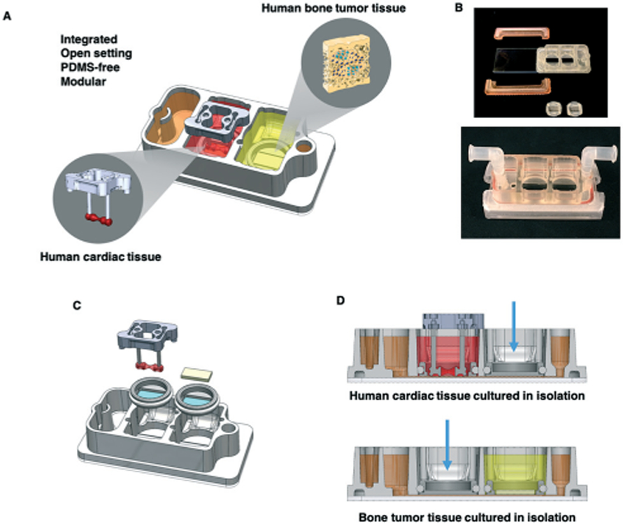
Fig. 1.

Experimental design. A. Schematic of the platform with two engineered human tissues: Ewing sarcoma (ES) tumor and cardiac tissues that were cultured either with microfluidic perfusion (integrated platform) or in isolation. Metastatic and non-metastatic ES tumors were studied at clinical dosages and treatment regimens of linsitinib. B. Photographs of the integrated platform and its components (top) and in its complete functional state (bottom). C. Platform assembly; note microfluidic connections for circulation at the left and right, and the reservoir for perfusate at the left. D. The platform setup for culturing tissues in isolation, as shown for the cardiac tissue (top) and the bone tumor tissue (bottom). Blue arrows indicate polypropylene plugs, allowing culture of one tissue at a time in isolation.
