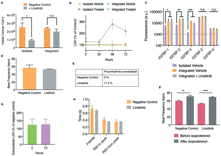
Fig. 5.

Responses of human engineered bone ES tumors and cardiac tissues to linsitinib in the integrated platform with microfluidic perfusion. A. and B. Non-metastatic ES bone tumors and cardiac tissues were exposed to linsitinib (12 μM) over a period of 72 hours in either isolated culture or within the perfused integrated platform. Luminescence (A) and LDH secretion (B) as functions of cancer cell number and viability as well as cytotoxicity, respectively, were measured (mean ± s.e.m., n = 3). C. Protein lysates were collected from non-metastatic ES bone tumors either grown in isolation, or exposed to perfusion and circulating linsitinib (12 μM) over a period of 72 hours in the integrated platform. Subsequently, comparative proteomic analysis of IGF-1 binding proteins was performed (mean ± s.d., n = 3 per group). D. Beat frequency of cardiac tissues after exposure to linsitinib (12 μM) within the perfused integrated platform (mean ± s.e.m., n = 9). E. Occurrence of proarrhythmic events/beat after exposure to linsitinib within the platform. F. Beat frequency of cardiac tissues that had been exposed to linsitinib in the platform after isoproterenol exposure (mean ± s.e.m., n = 9). G. Extracellular LDH before and after linsitinib exposure, as percentage of negative control (mean ± s.e.m. n = 3). H. Calcium transients of cardiac tissues characterized by the full-width half-maximum (FWHM), R50 to and from peak times (50% of the time to and from the maximal peak of the calcium transient) (mean ± s.e.m. n = 8–9). *P < 0.05; **P < 0.01; ***P < 0.001; ****P < 0.0001 by two-way ANOVA with Bonferroni post-test or unpaired, two-tailed Student's t test.
