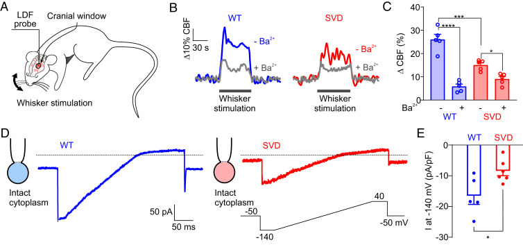
Fig. 2.
Kir2.1 channel down-regulation underlies deficits in capillary-to-arteriole electrical signaling in SVD. (A) Whisker stimulation experimental scheme. (B) Representative traces showing whisker stimulation-induced changes in CBF in control WT (blue) and SVD (red) mice in the presence (gray traces) and absence of Ba2+ (100 µM). (C) Summary data from five WT and five SVD mice (*P < 0.05, ***P < 0.001, ****P < 0.0001, repeated-measures two-way ANOVA). (D) Representative traces of Kir2.1 currents in cECs from WT (blue) or SVD (red) mice, recorded in the perforated-patch configuration. (E) Summary data showing Kir2.1 currents (n = 5–6 cECs from three mice per group; *P < 0.05, unpaired Student’s t test).

