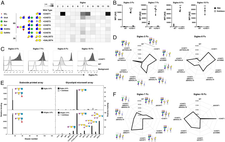
Fig. 5.
Probing the contribution of sulfotransferases to Siglec sialoglycan recognition. (A) Sulfotransferases knocked in into HEKWT cells and predicted sulfated glycan structures are shown. Heat map shows Siglec Fc binding to the KI cells as MFI values normalized to HEKWT. (B) Dot plots show Siglec-3/7/8/15 binding to HEKWT and HEKKI CHST1 cells treated with PBS or sialidase. Data of three independent experiments are presented as average MFI ± SEM. (C) Representative histograms show binding of Siglec-3/7/8/15 to CHOWT and CHOKI CHST1 cells. Cells stained only with anti-human IgG AF647 indicate background fluorescence. (D) Radar charts show Siglec-3 (Left) and Siglec-8 (Right) binding to HEKWT and HEKKI CHST1 cells with additional KO of O-GalNAc glycans (KO COSMC), N-glycans (KO MGAT1), glycolipids (KO B4GALT5), N-glycans and glycolipids (KO MGAT1, KO B4GALT5), or α2-3Sia on N-glycans (single or double KO ST3GAL4/6). (E) Siglec binding to glycan arrays. Recombinant Siglec-8 and Siglec-3 Fc chimera were tested for binding to two sialoglycan arrays: a 123-glycan array printed on glass slides (Left) and an 11-glycan sialoglycolipid array on 384-well plates. The glycan structures tested are listed in SI Appendix. Binding is expressed in arbitrary fluorescence units for the printed array and as colorimetric enzyme activity (ΔA405/min × 1,000) for the glycolipid array. Each point is the average of quadruplicate determinations. As indicated, a replicate glycolipid array was pretreated with 50 mU/mL Vibrio cholerae sialidase for 90 min at 37 °C and washed prior to addition of the Siglec Fc chimeras. (F) Radar chart shows Siglec-7 (Left) and Siglec-15 (Right) binding to HEKWT and HEKKI CHST1 cells with additional KO of O-GalNAc glycans (KO COSMC), core2 (KO GCNT1), N-glycans (KO MGAT1), glycolipids (KO B4GALT5), or N-glycans and glycolipids (KO MGAT1, KO B4GALT5). Representative MFI values of three independent experiments are shown, and deleted glycoconjugates are depicted in gray.

