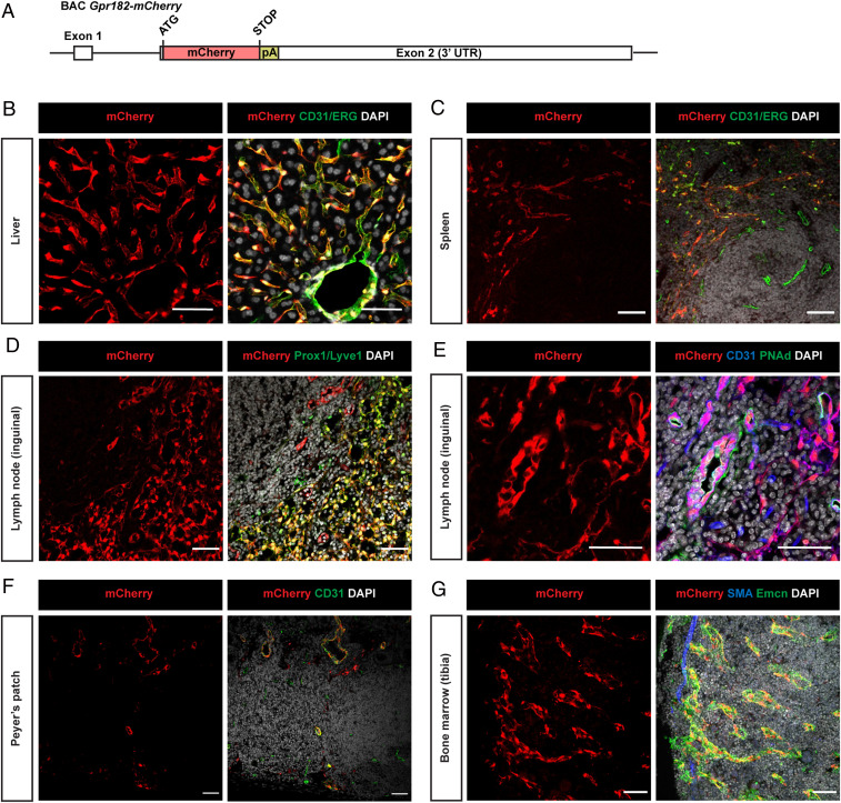
Fig. 1.
GPR182 is expressed in microvascular endothelial cells. (A) Schematic of part of the BAC-based mouse Gpr182-mCherry reporter transgene, which had a total length of 234 kb. UTR: untranslated region. (B–G) Representative immunofluorescence confocal images of cryosections of the indicated organs from Gpr182-mCherry BAC transgenic mice. The mCherry signal corresponds to endogenous mCherry fluorescence. Cryosections were stained with antibodies against vascular or lymphatic endothelial markers (CD31 and ETS-related gene (ERG), as well as Prox1 and Lyve1, respectively). PNAd antibody was used to specifically mark high endothelial venules from lymph nodes. mCherry was often predominantly localized in the nucleus. Shown are results of representative experiments of at least three independently performed experiments. (Scale bars, 50 µm.)

