Fig. 2.
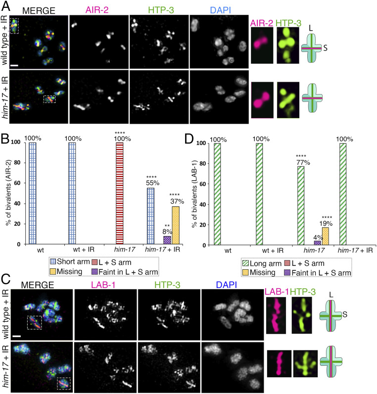
AIR-2 and LAB-1 localization on bivalents in oocytes at diakinesis in wild type and him-17 mutants after exogenous DSB formation. High-magnification images of diakinesis stage oocytes in wild type and him-17 mutant after exogenous DSB formation by γ-IR (60 Gy). (A) Immunolocalization of AIR-2 (magenta), HTP-3 (green), and DAPI (blue) in -1 oocytes at diakinesis indicates that AIR-2 is restricted to the short arm of the bivalents in him-17 mutants, similar to wild type, after IR treatment. A total of 106 and 88 bivalents from the gonad arms of 20 and 16 animals were analyzed for wild type and him-17, respectively, from three biological replicates. Dashed boxes indicate the bivalents for which AIR-2 and HTP-3 localization are shown at higher magnification. (Middle) The chromosome configuration observed at this stage showing distinct short (S) and long (L) arms, and the localization of AIR-2 (magenta) and HTP-3 (green) for each genotype following IR treatment. (B) Histogram indicates the percentage of bivalents at diakinesis with AIR-2 restricted to the short arm, mislocalized to both long and short arms, faint but on both long and short arms, or missing from the chromosomes in wild type and him-17 mutants with and without IR treatment. (C) Immunolocalization of LAB-1 (magenta), HTP-3 (green), and DAPI (blue) in -1 oocytes at diakinesis. A total of 91 and 103 bivalents from the gonad arms of 16 and 18 animals were analyzed for wild type and him-17, respectively, from three biological replicates. (D) Histogram indicates the percentage of bivalents at diakinesis with LAB-1 restricted to the long arms, mislocalized to both long and short arms, faint but on both long and short arms, or missing from the chromosomes in wild type and him-17 mutants with and without IR. ****P < 0.0001, **P < 0.005 by the two-tailed Fisher’s exact test. (Scale bars, 2 μm.) For complete/additional statistical analysis, see Dataset S1.

