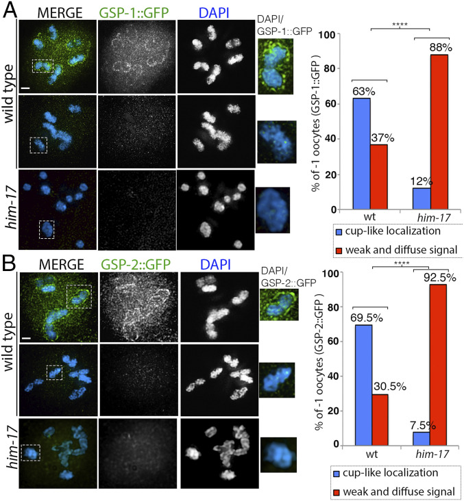
Fig. 5.
GSP-1::GFP and GSP-2::GFP expression in wild type and him-17 mutant. (A) High-magnification images of diakinesis stage -1 oocytes from animals expressing GSP-1::GFP stained with anti-GFP (green) and DAPI (blue) in wild type and him-17 mutant backgrounds. Dashed boxes indicate the bivalents shown at higher magnification to the right for clearer visualization of either the presence or absence of GSP-1::GFP cup-like localization. (Right) Histogram indicates the percentage of -1 oocytes with cup-like localization of GSP-1::GFP on the chromosomes (blue) or a weak and diffuse signal not on the chromosomes (red) in wild type and him-17 mutant. A total of 30 and 33 -1 oocytes from 30 and 33 animals were scored for wild type and him-17, respectively, from three biological repeats. (B) High-magnification images of diakinesis stage -1 oocytes from animals expressing GSP-2::GFP stained with anti-GFP (green) and DAPI (blue) in wild type and him-17 mutants. Dashed boxes indicate the bivalents shown at higher magnification to the right for clearer visualization of either the presence or absence of GSP-2::GFP cup-like localization. (Right) Histogram indicates the percentage of -1 oocytes with cup-like localization of GSP-2::GFP on the chromosomes (blue) or weak and diffuse signal not on the chromosomes (red) in wild type and him-17 mutants. A total of 23 and 27 -1 oocytes from 23 and 27 animals were scored for wild type and him-17, respectively. ****P < 0.0001 two-tailed Fisher’s exact test. (Scale bars, 2 μm.)

