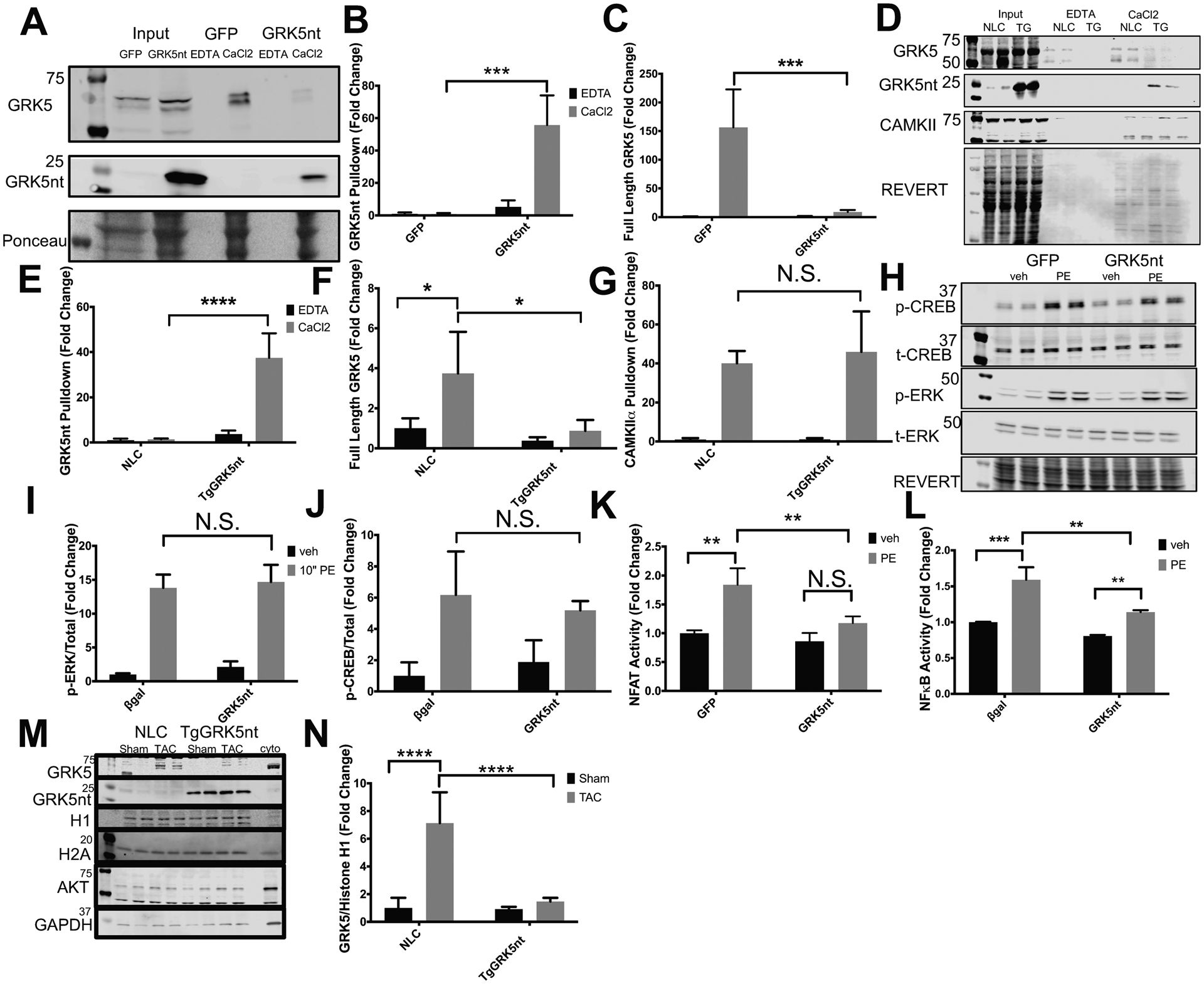
Fig. 5. GRK5nt binds to Ca2+-CaM selectively and alters NFAT signaling and GRK5 nuclear accumulation following hypertrophic stress.

(A) Western blot of GFP and GRK5nt lysates with CaM pulldown in the presence of EDTA or CaCl2. (B and C) Quantification of CaM pulldown of GRK5nt (B) or full-length GRK5 (C). (D) Western blot of CaM pulldown in NLC and TgGRK5nt lysates. (E–G) Quantification of CaM pulldown of GRK5nt (E), full-length GRK5 (F), or CAMKII (G). (H) Western blot of response to 10 min of PE stimulation in NRVMs expressing GFP or GRK5nt. Quantification of phospho-ERK (I) and phospho-CREB (J). Assessment of NFAT (K) or NFkB (L) luciferase activity in GFP or GRK5nt-expressing NRVMs following 24 hrs of 100μM PE treatment. (M–N) Western blotting (M) and quantification (N) of nuclear GRK5 in fractions from NLC or TgGRK5nt hearts two weeks after TAC. Data were normalized to the nuclear marker Histone H1. N=3 lysates per infection condition from independent cell isolations for (A–C). N=3 NLC heart lysates and N=4 TgGRK5nt heart lysates for (D–G). N=3 independent cell isolations for (H–L). N=4 NLC Sham mice, N=5 NLC TAC mice, N=4 TgGRK5nt Sham mice, N=4 TgGRK5nt TAC mice for (M–N). (* p<0.05, ** p<0.01, *** p<0.001, **** p<0.0001, two-way ANOVA with Tukey’s post-hoc testing).
