
Figure 3 |. The micrographs of tissue sections from control (a, c, e, g) or PEXTKO animals (b, d, f, h) stained with hematoxylin and eosin.
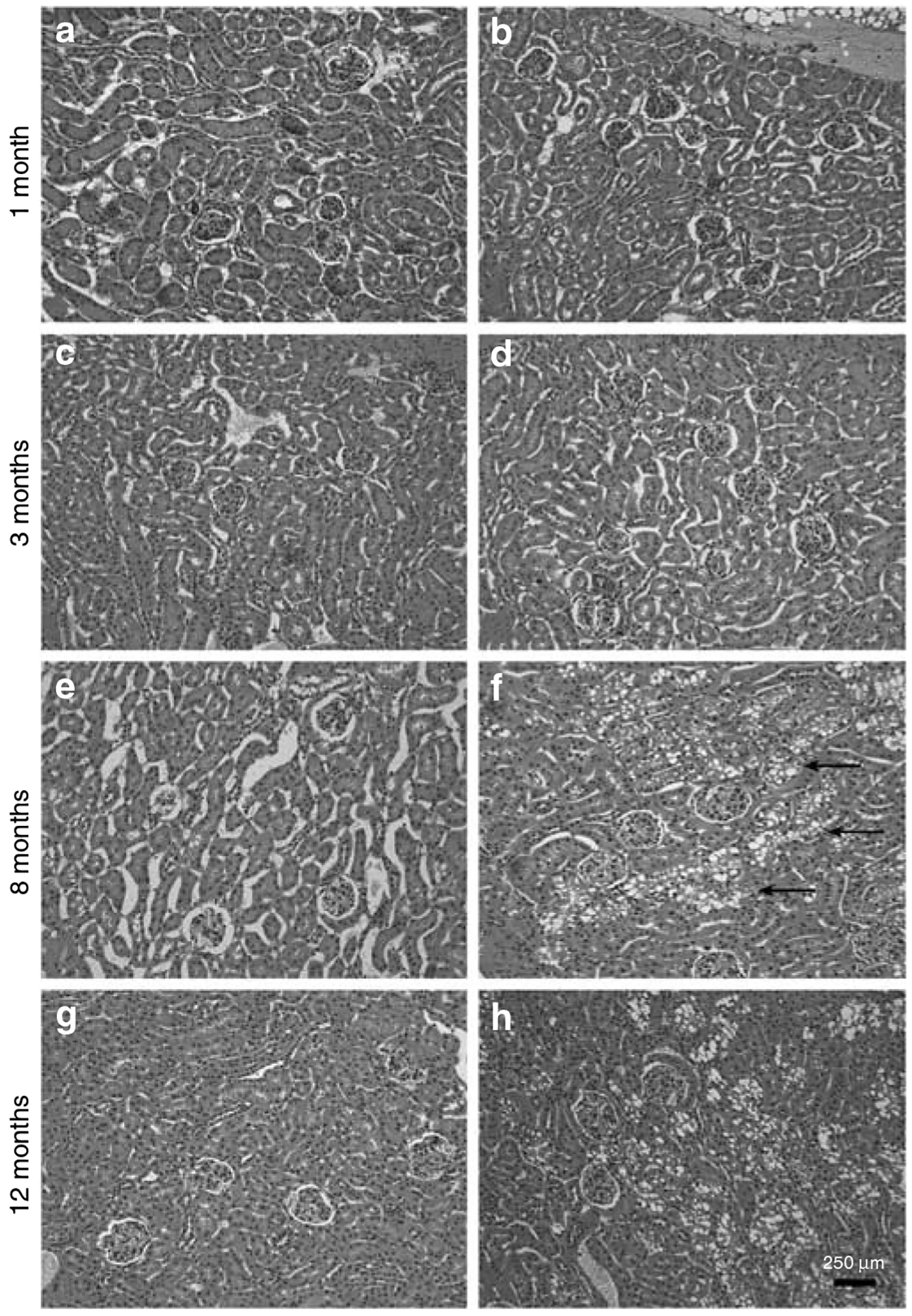
The age-matched animals were 1 (a, b), 3 (c, d), 8 (e, f), and 12 (g, h) months of age. Overall there were no profound differences in either glomerular or tubular structure between control and PEXTKO kidney at 1 and 3 months of age; no differences among the control animals were seen at any age examined. At 8 months of age, there were very apparent changes in the proximal tubule epithelial cells in the PEXTKO animals (f, arrows), the cells of which had large vacuoles present in the cytoplasm. These changes persisted in older PEXTKO animals (h).