
Figure 6 |. The electron micrographs show glomeruli from adult control animals (a, 2.5P-Cre+/Ext1wt/wt) and PEXTKO (b, 2.5P-Cre+/Ext1fl/fl) animals.
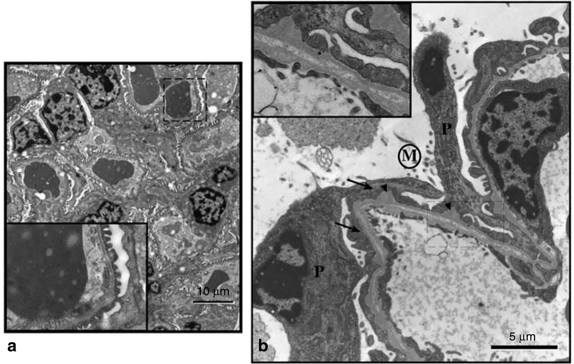
In control animals, the basic architecture of the glomeruli from these animals is normal, with no visible sign of foot process effacement or obvious BM ‘outpockets.’ The inset in the corner of each larger micrograph is a higher magnification of the area in the main micrograph outlined by the dashed line. (b) An electron micrograph of a glomerular capillary wall from a 1-month-old PEXTKO mouse. Within the picture are the cell bodies of two podocytes (P) having abnormal, effaced foot processes (arrows). On the apical surface of the podocytes, microvilli (area denoted by M) can be found. The GBM also shows irregularities, having numerous outpockets or ‘humps’ (arrowheads) on the podocyte side of the GBM. The inset shows an enlargement of the boxed-in area of the capillary wall. In this region, the BM has an abnormal appearance, resembling two separate BMs. The presence of the outpockets, along with the double BM, suggests the possibility that GBM fusion, a normal developmental process, has been delayed in this area of the glomerulus. The presence of a double BM has not been seen in older (>3 months) animals.