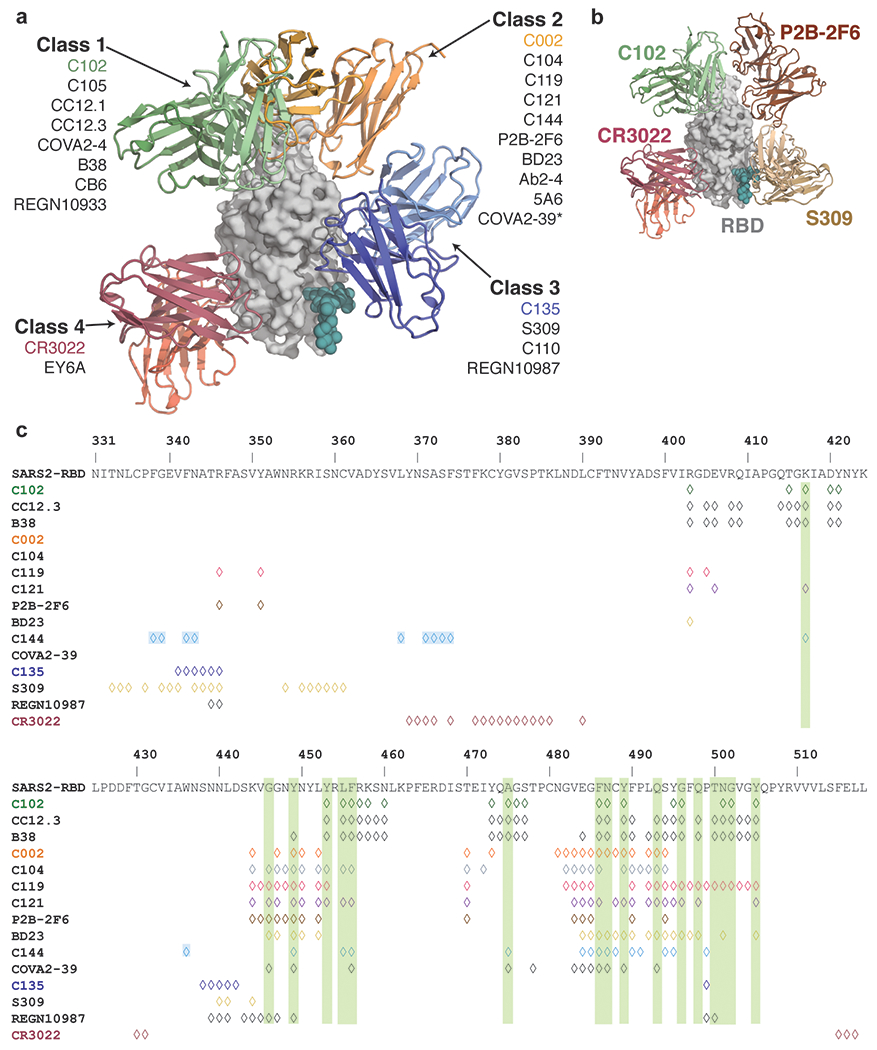
Extended Data Figure 9: Summary of hNAbs.

a, Structural depiction of a representative NAb from each class binding its RBD epitope. b, Composite model illustrating non-overlapping epitopes of NAbs from each class bound to a RBD monomer. c, Epitopes for SARS-CoV-2 NAbs. RBD residues involved in ACE2 binding are boxed in green. Diamonds represent RBD residues contacted by the indicated antibody.
