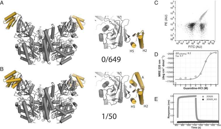
Fig. 2.
Design of a hTfR binding protein. (A) Model of first generation TfR binders (gray: TfR ectodomain, yellow: binder). (B) Model of the second generation TfR binders (gray: TfR ectodomain, yellow: binder). (C) 2DS25 (design: gray, negative control: black) binds to hTfR ectodomain in flow cytometry. A total of 100,000 cells were measured. (D) CD chemical denaturation experiment of 2DS25. (E) Single concentration biolayer interferometry assay (gray: 2DS25, black: 2DS25_KO [W81A/Q85A]).

