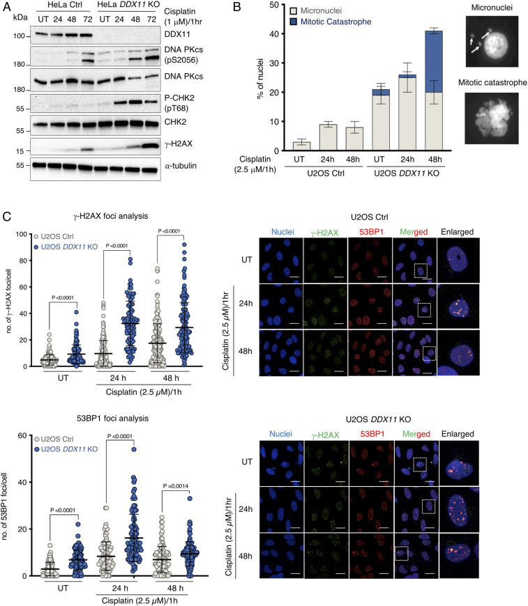
Fig. 2.
DDX11 loss associates with persistent DNA damage accumulation and micronucleation. (A) HeLa Ctrl and DDX11 KO cells were treated with cisplatin (1 μM) for 1 h and allowed to recover for 72 h, during which DNA damage markers were analyzed by Western blotting at the indicated time points (n = 2). (B) Quantification of micronuclei and mitotic catastrophes in U2OS Ctrl and DDX11 KO cells in untreated conditions and upon recovery from an acute cisplatin treatment (2.5 μM for 1 h). Error bars show average ± SEM. (C) Quantification and representative micrographs of γ-H2AX and 53BP1 foci in U2OS Ctrl and DDX11 KO cells recovering from an acute treatment with cisplatin (2.5 μM for 1 h). (Scale bar, 10 μm.) n = 2. Statistical analysis of foci was performed using Student’s t test. Error bars show average ± SD.

