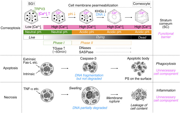
Fig. 5.
A schematic illustration of the hypothetical cell death process of corneoptosis. In “corneoptosis,” corneocytes are formed after SG1 cell death through two sequential, intracellular states. “phase I:” high [Ca2+]i-neutral pHi activates TGases and lasts for ∼60 min. “phase II:” high [Ca2+]i-acidic pHi induces the activation of DNases (DNase1L1 and DNase2), SASPase, and changes in the keratin network. As a result, KHGs are eliminated, the DNA is completely degraded, then the process of SG1 cell death is completed (black shaded bar), and corneocytes are formed. In apoptosis, extrinsic (Fas-L) or intrinsic factors activate the apoptotic pathway via the activation of effector caspases (such as caspase-3). Activated DNases fragment DNAs but do not degrade them completely. Organelles are degraded. Cytochrome C is released from the mitochondria. Chromatin is condensed. Cells finally form apoptotic bodies that present phosphatidylserine (PS) on their surface. PS is recognized by macrophages or adjacent cells that phagocytize dead cells and degrade all dead cell components. In necrosis, dead cells become swollen, and organelles/DNAs are partially degraded; subsequently, cell membranes are finally ruptured, and cell contents are leaked. This causes immune responses resulting in inflammation and the removal of cell remnants by immune cells via efferocytosis.

