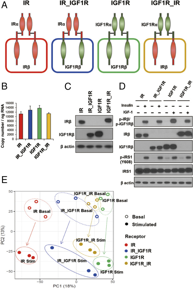
Fig. 1.
Phosphoproteomic analysis of cells expressing IR, IGF1R, and the chimeric receptors IR_IGF1R and IGF1R_IR. (A) Schematic representation of the experimental strategy applied to analyze the phosphoproteome of DKO preadipocytes reconstituted with IR, IGF1R, and chimeric receptors IR_IGF1R and IGF1R_IR. (B) Relative mRNA levels of recombinant receptors of IR, IGF1R, IR_IGF1R, and IGF1R_IR as determined by qPCR using cDNA standards for quantitation. Data are means ± SEM copy number per nanogram of total RNA, n = 3. (C) Immunoblotting of IR and IGF1R using antibodies specific for IRβ (#3025, Cell Signaling) and IGF1Rβ (#3027, Cell Signaling) in lysates from cells expressing IR, IGF1R, IR_IGF1R, and IGF1R_IR receptor cDNAs. (D) Immunoblotting to determine phosphorylated and total receptor protein levels and IRS-1Y608 phosphorylation in lysates from IR and chimeric receptor IR_IGF1R stimulated with 10 nM insulin or IGF1R and chimeric receptor IGF1R_IR stimulated with 10 nM IGF1 for 15 min. (E) PCA of the phosphosites identified by LC-MS/MS from three independent lines of DKO preadipocytes reconstituted with IR, IGF1R, and chimeric receptors IR_IGF1R and IGF1R_IR in the basal and hormone-stimulated states. Cells were serum starved for 5 h with DMEM containing 0.1% BSA before insulin and IGF1 stimulation.

