FIG 4.
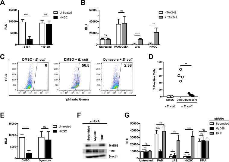
LPS- and GC-mediated repression of HIV-1 replication in MDMs requires TLR4, TRIF, and type I IFNs. (A) MDMs were infected with a single-round, replication-defective HIV-luciferase reporter virus and, 48 h after infection, cells were treated with GC (MOI = 10) in the absence (white bars) or presence (black bars) of B18R (100 ng/ml) for 18 h. The cells were then lysed and assayed for luciferase activity. The data are the mean (± SD) of seven donors; each donor was tested in triplicate. (B) MDMs were infected as in panel A and, 48 h after infection, were treated with PAM3CSK4 (100 ng/ml), LPS (100 ng/ml), or heat-killed GC (MOI = 10) in the absence (white bars) or presence (gray bars) of TAK242 (1 μg/ml) for 18 h. The cells were then lysed and assayed for luciferase activity. The data are the mean (± SD) of six donors; each donor was tested in triplicate. (C and D) MDMs were incubated with dimethyl sulfoxide (DMSO) or the dynamin inhibitor Dynasore (80 μM) for 15 min at 37°C. The cells were washed with phosphate-buffered saline (PBS) and incubated with pHrodo green E. coli (1 mg/ml) for 2 h at 37°C. Endocytosis/phagocytosis was measured by flow cytometry. Shown are data from one representative donor (C) and composite data from four donors (D). (E) MDMs were infected as in panel A and, 48 h after infection, were treated with vehicle control (white bars) or with Dynasore (80 μM); black bars for 15 min prior to treatment with heat-killed GC (MOI = 10) for 18 h. The cells were then lysed and assayed for luciferase activity. The data are the mean (± SD) of six donors, each donor tested in triplicate. (F and G) MDMs were transfected with a control scrambled shRNA, shRNA targeting MyD88, or shRNA targeting TRIF. Knockdown of protein expression was detected by Western blot analysis (F). Transfected MDMs were infected with a single-round, replication-defective HIV-luciferase reporter virus and, 48 h after infection, were treated with PAM3CSK4 (100 ng/ml), LPS (100 ng/ml), heat-killed GC (MOI = 10), or phorbol myristate acetate (PMA) (10 nM) for 18 h. The cells were then lysed and assayed for luciferase activity (G). The data are the mean (± SD) of six donors. *, P < 0.05; **, P < 0.01; ***, P < 0.001; ****, P < 0.0001; ns, not significant.

