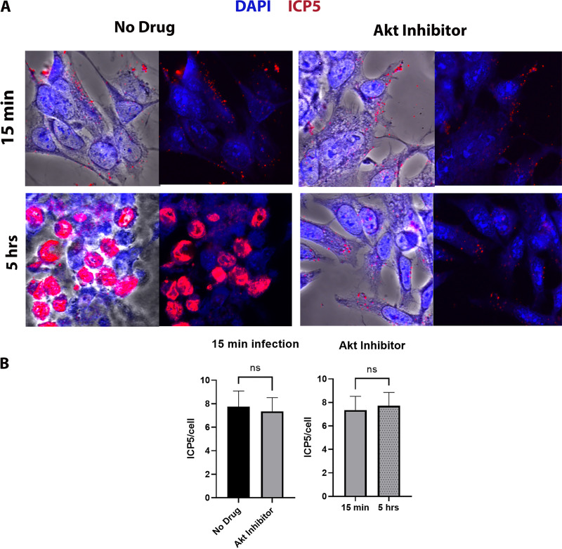
FIG 1.
Effect of Akt HSV-1 transport. (A) SK-N-SH cells were adsorbed at 4°C with HSV-1 (McKrae) at an MOI of 20 for 1 h, shifted to 37°C for 15 min, and then washed with low-pH buffer. The Akt inhibitor miltefosine was added at a concentration of 30 μM, and the mixture was incubated for 5 h at 37°C. The 8-well chamber slide was then fixed with formalin, permeabilized with methanol, and prepared for fluorescence microscopy. Antibody against VP5 (ICP5) (red) was used, and nuclei were stained with DAPI (blue). Capsids colocalized in the nuclei appear purple. Magnification is 63× under oil immersion. (B) The amount of fluorescence detected with the anti-ICP5 antibody (virion capsids) was quantified by fluorescence imaging of 10 different random sections of slides, using ImageJ software. The fluorescence signals per cell were counted, and their average value was used to plot the graph. Statistical comparison was conducted by Graph Pad Prism using Student's t test. Error bars represent the 95% confidence interval about the mean. Differences were determined significant at P < 0.05.

