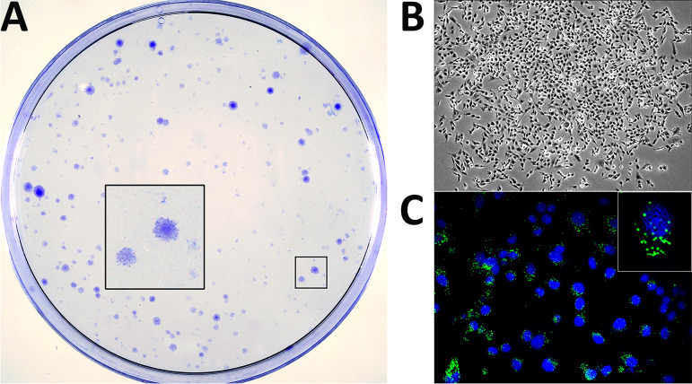
FIG 1.
Generation of persistently infected DF-1 cell cultures. DF-1 cell monolayers were infected with IBDV at an MOI of 3 PFU/cell. At 4 days p.i., cultures were carefully rinsed to remove cell debris and then maintained in DMEM supplemented with 10% FCS. Culture medium was replaced every 5 days. At 21 days p.i., cultures were either stained with crystal violet to visualize surviving cell clones (A) or processed for phase-contrast (B) or IF (C) microscopy after incubation with an antibody specifically recognizing the IBDV structural VP3 polypeptide (green). Cell nuclei (blue) were stained with DAPI. Insets show a higher magnification (×2.5) of boxed areas.

