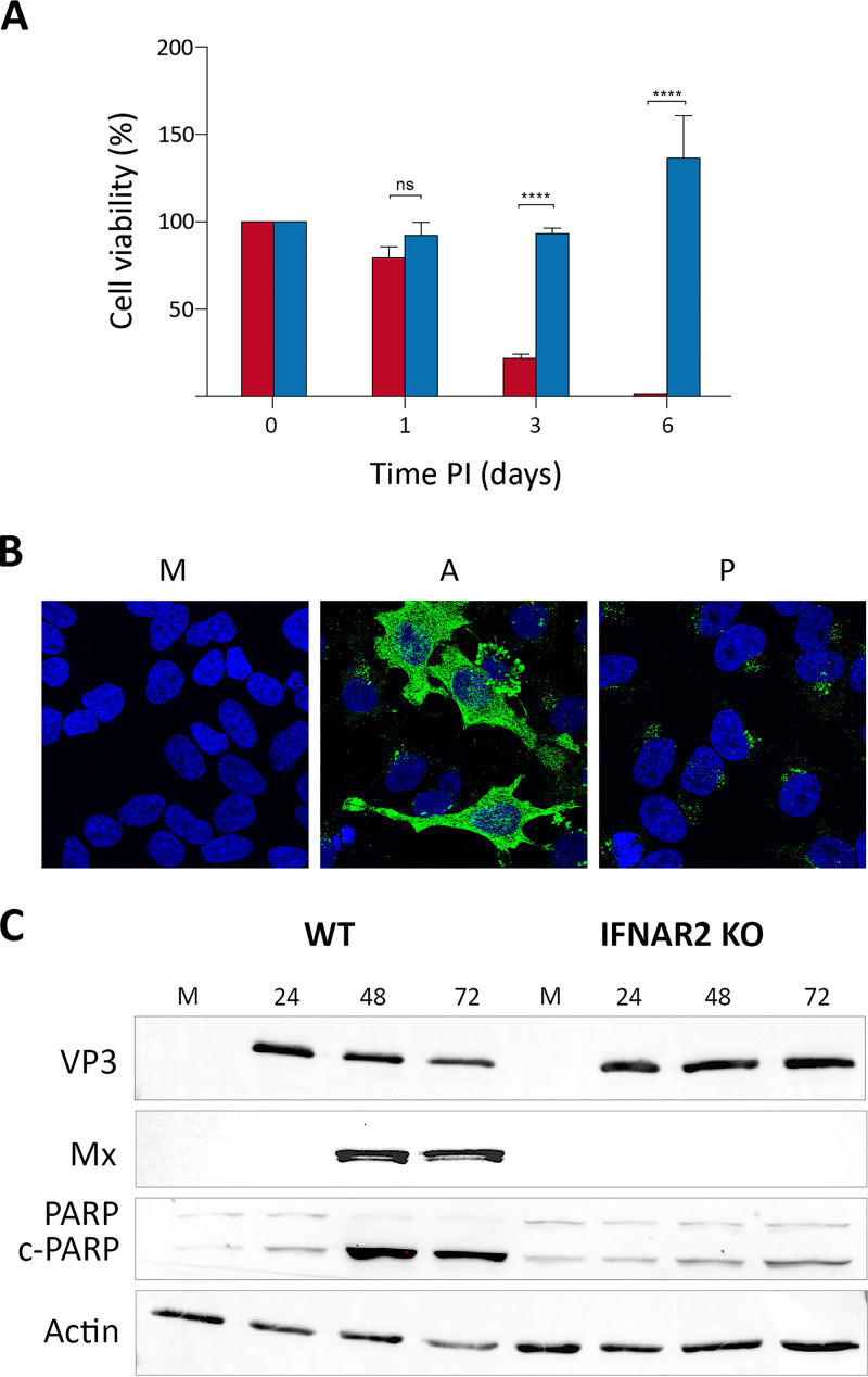
FIG 7.
Effect of the inactivation of the JAK-STAT pathway on the fate of IBDV-infected HeLa cells. (A) WT (red) and IFNAR2 KO HeLa (blue) cell cultures were infected with WT IBDV (3 PFU/cell). Cell viability was determined at the indicated times p.i. using the MTT assay. MTT values recorded immediately after infection were considered 100% cell viability. Presented data correspond to the means ± standard deviations from three independent experiments. Brackets indicate pairwise data comparisons. ****, P < 0.00001 as determined by two-way ANOVA. ns, not significant. (B) Persistently infected IFNAR2 KO cells (P) maintained for 2 months were processed for IF analysis using an antibody specifically recognizing the IBDV structural VP3 polypeptide. Cell nuclei (blue) were stained with DAPI. Mock-infected (M) and acutely infected (3 PFU/cell, fixed at 48 h p.i.) (A) cells were used as controls. (C) Infected (3 PFU/cell) WT and IFNAR2 KO cell cultures were collected at the indicated times p.i. Mock-infected (M) cell cultures were used as controls. The corresponding extracts were subjected to SDS-PAGE followed by Western blotting using antibodies against the virus-encoded VP3 and the cellular Mx, PARP, and actin proteins. The PARP cleavage product is denoted c-PARP.

