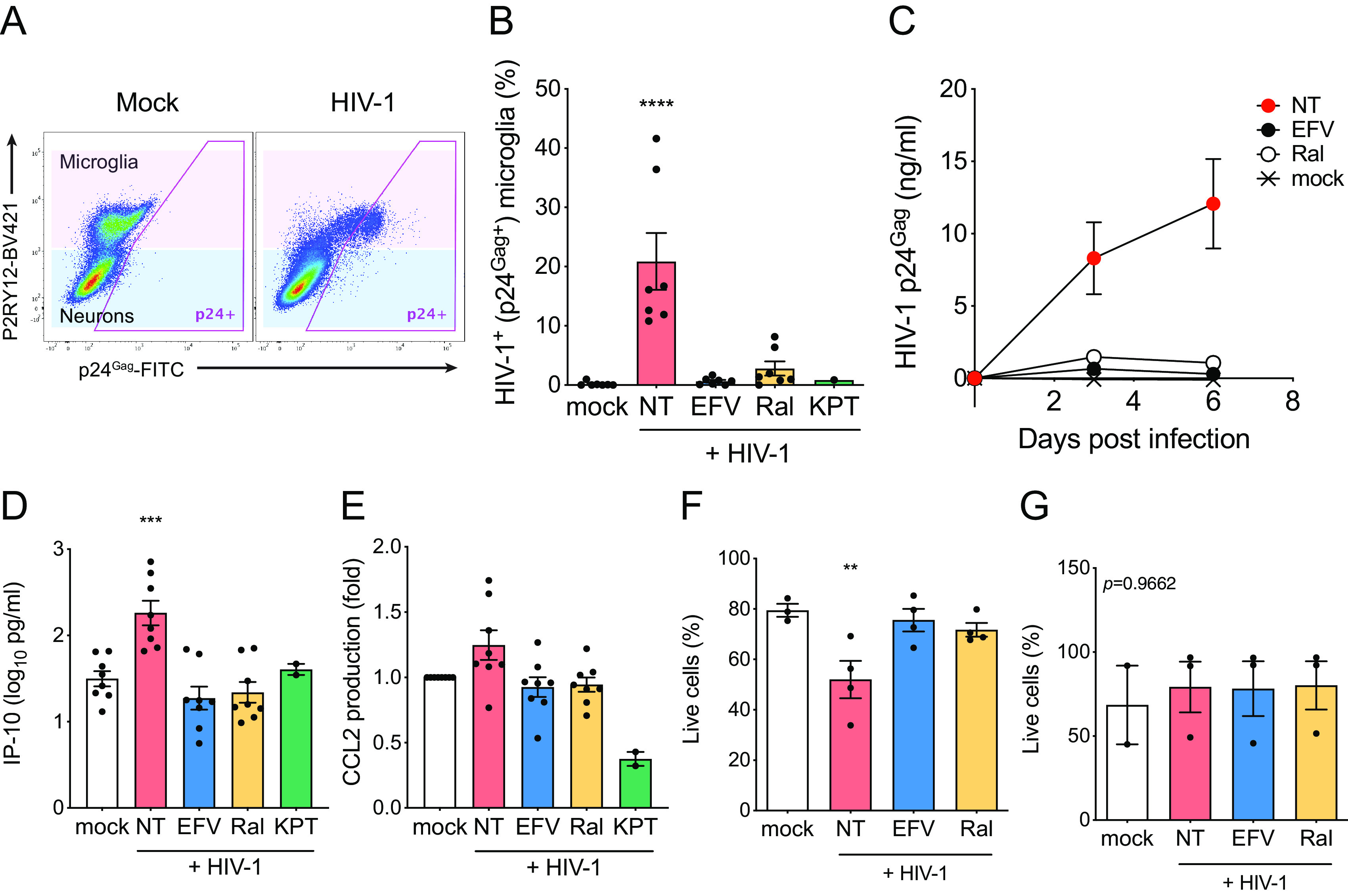
FIG 5.

HIV-1 infection of hiMGs in hiMG-hiNeuron cocultures induces proinflammatory responses. hiMG-hiNeuron cocultures were infected with HIV-1 (Lai/YU-2env: replication-competent CCR5 tropic HIV-1, MOI = 1). (A and B) HIV-1 infection (intracellular p24Gag expression) was analyzed by flow cytometry. (A) Representative flow cytometry profile is shown, and microglia (P2RY12+) and neuron (P2RY12−) populations are highlighted with pink and blue, respectively. (B) HIV-infected (p24Gag+) cells in microglia (pink in panel A) were calculated. (C) Replication kinetics of HIV-1 in hiMG-hiNeuron coculture. Cocultures were infected with HIV-1 (Lai/YU-2env, replication-competent CCR5 tropic HIV-1, MOI = 1), and production of p24Gag in the culture supernatant was quantified by ELISA. (D and E) Production of proinflammatory cytokines (D) IP-10 and (E) CCL2 was measured by ELISA (6 dpi). (F and G) The proportion of live cells in (F) microglia (pink in panel A) and (G) neurons (blue in panel A) was calculated. The means ± SEM are shown, and each symbol represents an independent experiment. P values: one-way ANOVA followed by Dunnett’s posttest comparing to mock (panels B and D to F); **, P < 0.01; ***, P < 0.001; ****, P < 0.0001. The P value from one-way ANOVA test was 0.9662 for panel G. NT, no-treatment (DMSO); EFV, efavirenz (1 µM); Ral, raltegravir (30 µM); KPT, KPT-330 (selinexor, 1 µM).
