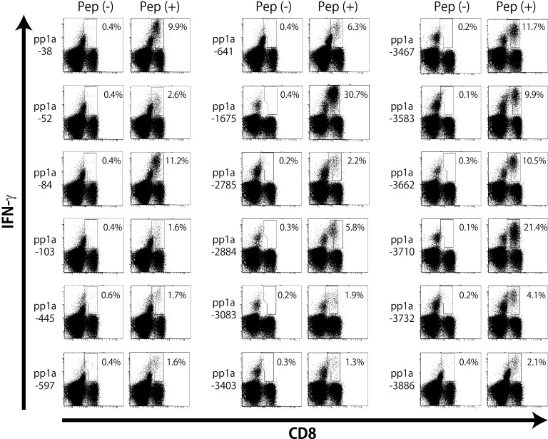
FIG 2.
Intracellular IFN-γ staining of CD8+ T cells specific for peptides derived from SARS-CoV-2 pp1a. HHD mice were immunized twice with each of the predicted peptides of SARS-CoV-2 pp1a in liposomes together with CpG. After 1 week, spleen cells were prepared and stimulated with (+) or without (−) a relevant peptide for 5 h. Cells were then stained for their surface expression of CD8 (x axis) and their intracellular expression of IFN-γ (y axis). The numbers shown indicate the percentages of intracellular IFN-γ+ cells within CD8+ T cells. The data shown are representative of three independent experiments. Three to five mice per group were used in each experiment, and the spleen cells of the mice per group were pooled.

