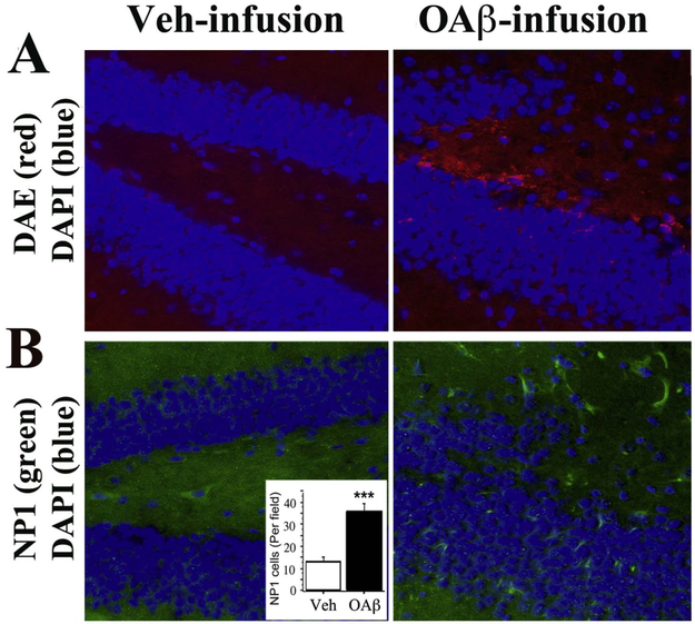
Fig. 5. Aβ42 oligomers infusion increases brain NP1.
After infusing 50 μg of Aβ42 oligomers (OAβ) into right ventricle of 4-month-old wile type (WT) mice over 2 weeks, brain adjacent serial sections were used for Aβ and NP1 staining. A. Aβ Deposition (red) was observed by immunostaining with anti-Aβ antibody, DAE, in the dentate gyrus of the hippocampus in OAβ-infused WT mice but not in the vehicle (Veh)-infused mice. B. NP1 (green) was increased in the dentate gyrus of the hippocampus in OAβ-infused WT mice but not in the Veh-infused mice. Quantification of immunostained NP1 cells confirmed a significant difference between the two groups (p < 0.001).

