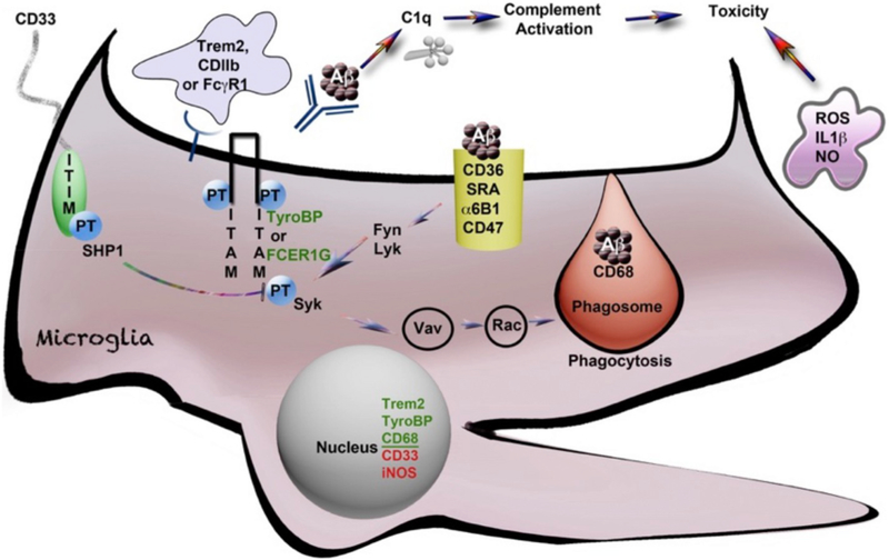
Fig. 1.
Activation of microglia and phagocytosis controlled by tyrosine phosphorylated AD risk-associated genes (*). Schematic diagram of a microglial cell with the Fc(g)R1 receptor for antibody, opsonized Aβ and parallel signaling receptors: TREM2* and CD11b*, all linked to ITAM domain phosphorylation signaling on TYROBP/DAP12* or FCER1G, that then signal through Syk tyrosine kinase to promote phagocytosis. This ITAM signaling is negatively regulated by CD33* (and other Siglecs with ITIM domains), which couple to SHPS like SHP1 (tyrosine phosphatases). SHP1 dephosphorylates (deactivates) the TYROBP/ITAM-activated, phagocytosis-promoting signaling pathways as shown. The activation of these signaling pathways is represented by phosphorylated tyrosine residues (“PT”). The process of phagocytic engulfment and a mature phagosome fusing with a lysosome (marked by the glycoprotein CD68* (mouse macrosialin) is shown with engulfed Aβ. The classic microglia marker CD68 is a lysosomal innate immune marker of the phagocytic microglial phenotype (reviewed in Zotova et al., 2011). CD11b (ITGAM) is a classic marker of M1 activation that complexes with CD18 (ITGB2), a proximal TyroBP partner. CD11b also serves as a receptor for complement C3b opsonized Aβ aggregates (Wyss-Coray and Mucke, 2002). C1q interacts directly with Aβ, upstream of C3, and its expression is markedly elevated in AD (Yasojima et al., 1999). This figure is adapted from Zhang and Painter (Painter et al., 2015; Zhang et al., 2013).

